craiyon logo
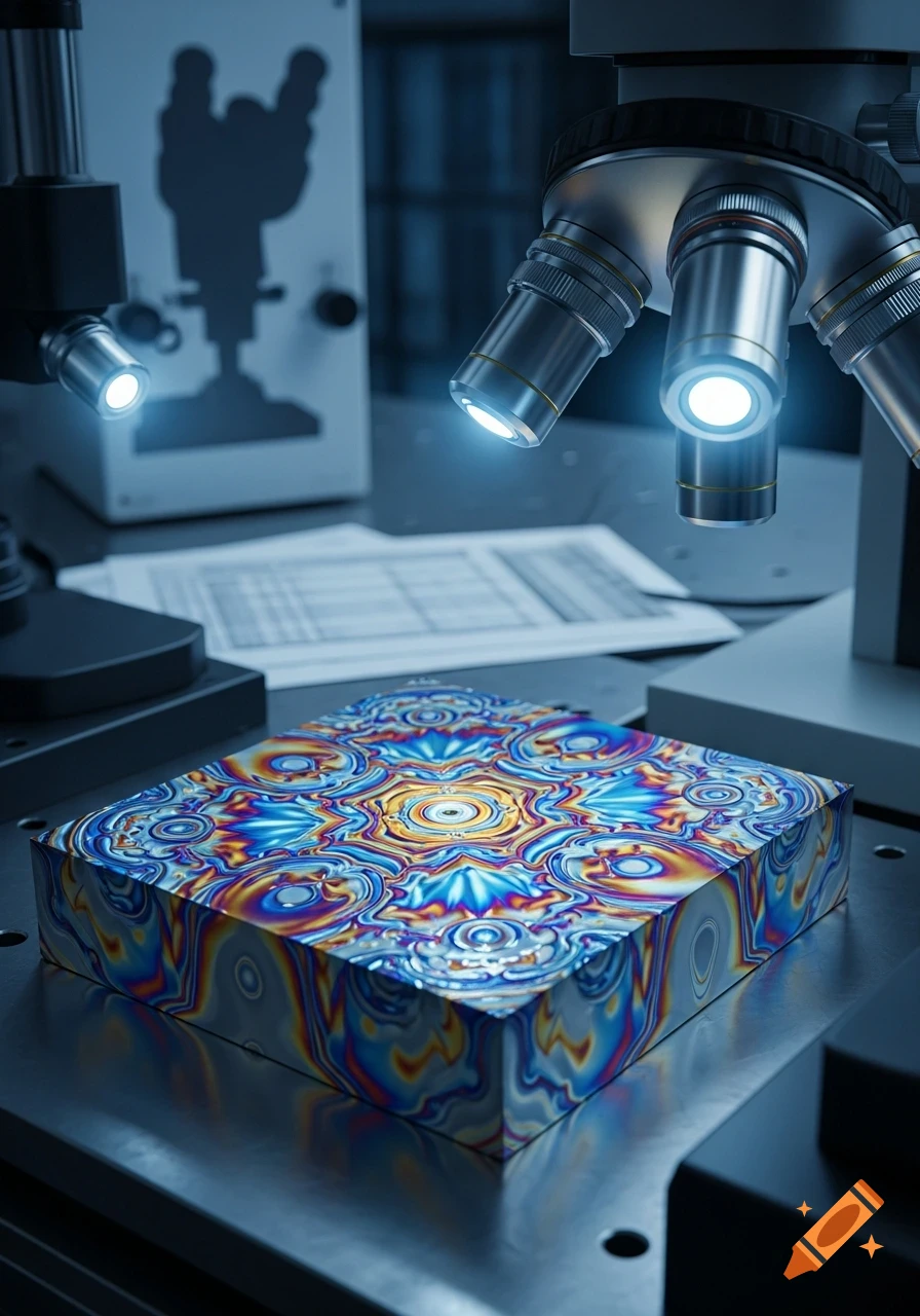
A close-up, photorealistic shot of a silver microscope examining a textured metal sample on a lab bench with notes and equipment.
Zoom-Aktion
Detailed microscopic view of a single red blood cell with a bright highlight, surrounded by other blurred red blood cells in a reddish fluid.
Zoom-Aktion
A close-up, photorealistic view of numerous red blood cells flowing through a transparent blood vessel.
Zoom-Aktion
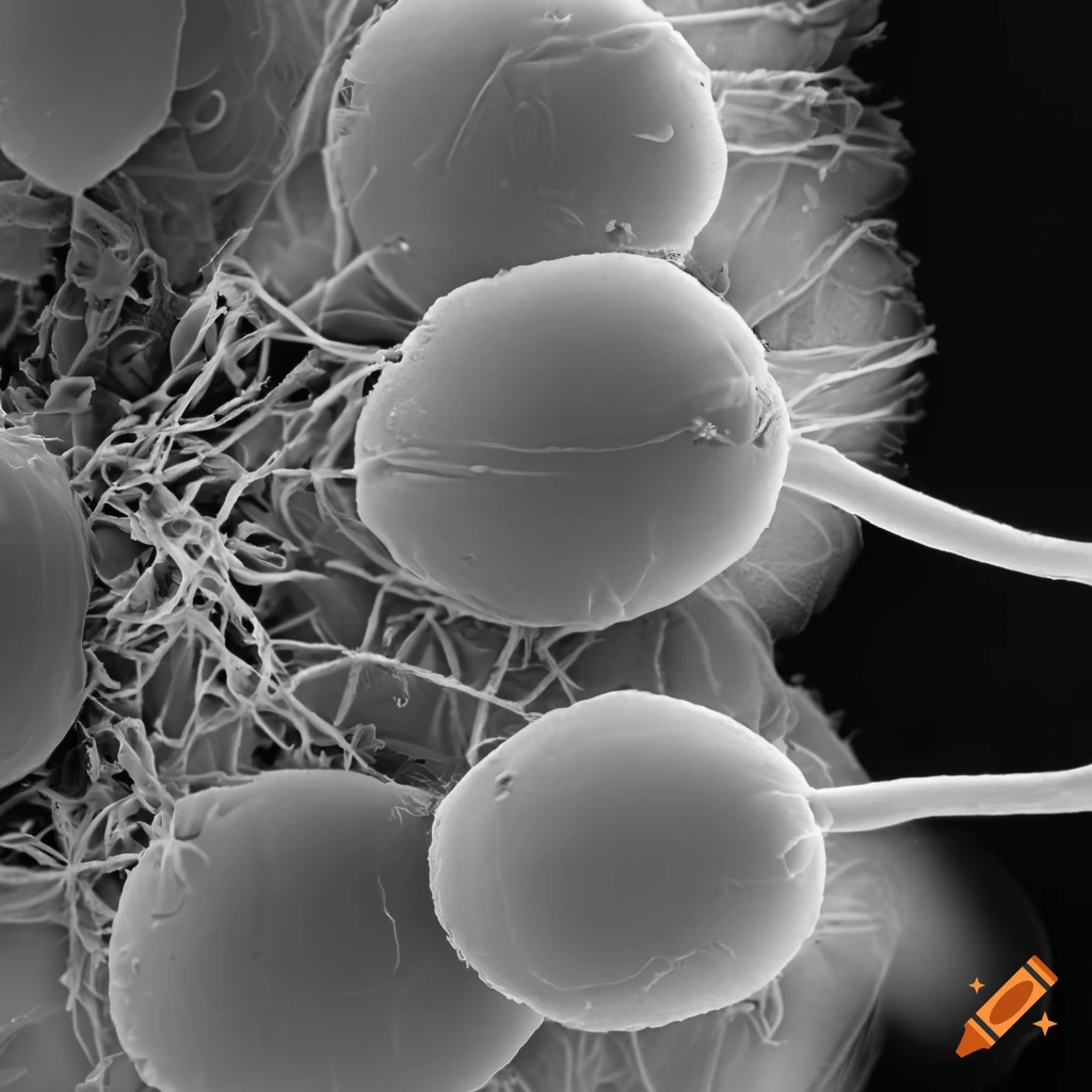
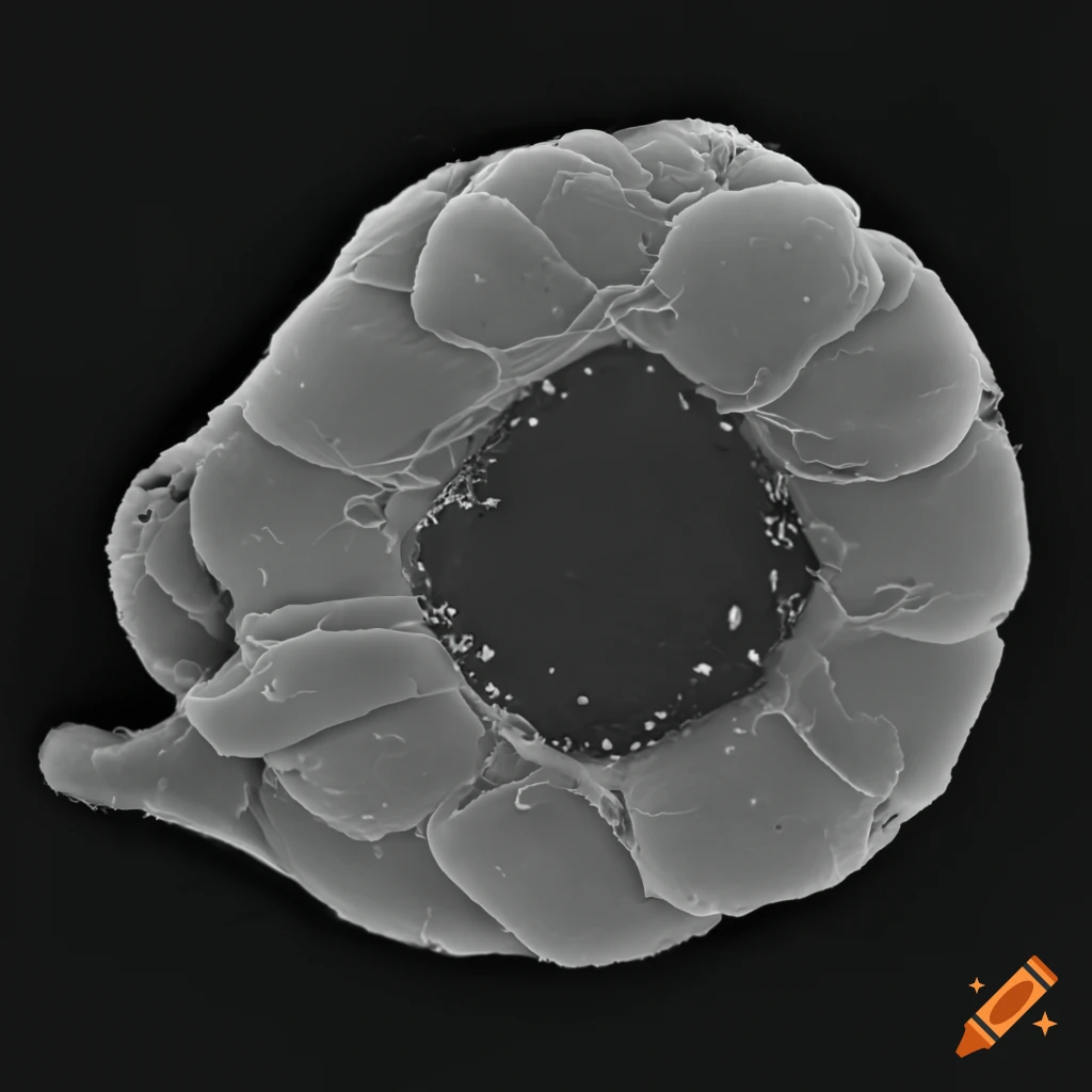
A detailed grayscale electron micrograph of a spherical particle covered in numerous small, rounded protrusions.
Zoom-Aktion
Photorealistic close-up of translucent, elongated soil bacteria with fine tendrils, nestled among brown soil clumps and pebbles.
Zoom-Aktion
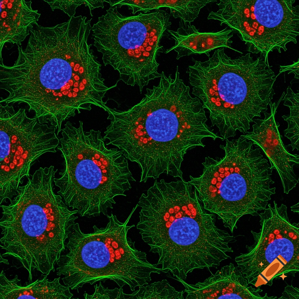
Immunofluorescence image of a cell with a blue nucleus, red golgi dots, and green actin filaments against a black background.
Zoom-Aktion
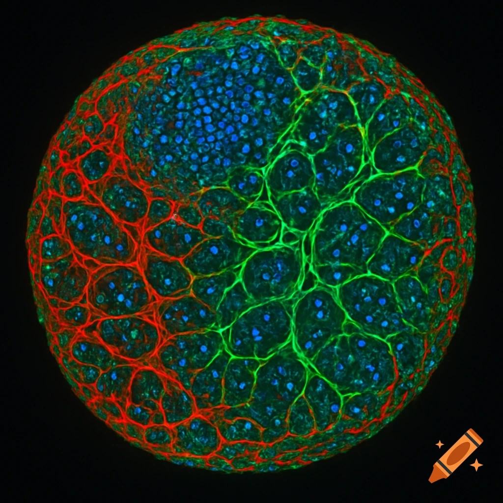
Fluorescence microscopy image of cells with blue nuclei, red microtubules, and green actin in a swirling 'eye of the storm' pattern on a black background.
Zoom-Aktion
Magnified view of a blue, textured cell with internal bubbles, resting on a beige fibrous tissue.
Zoom-Aktion
A detailed, microscopic view of a neuron or dendritic cell, featuring a central purplish-blue cell body with numerous branching extensions, against a soft green and yellow bokeh background.
Zoom-Aktion
A purple bacteriophage with long, thin legs attaching to a large, textured brown cell, depicted in a detailed microscopic view.
Zoom-Aktion
Close-up, detailed microscopic view of a human sperm cell with a clear head and tail on a white background, photorealistic.
Zoom-Aktion
Detailed 3D illustration of multiple green chloroplasts with internal thylakoid stacks, floating in a microscopic view.
Zoom-Aktion
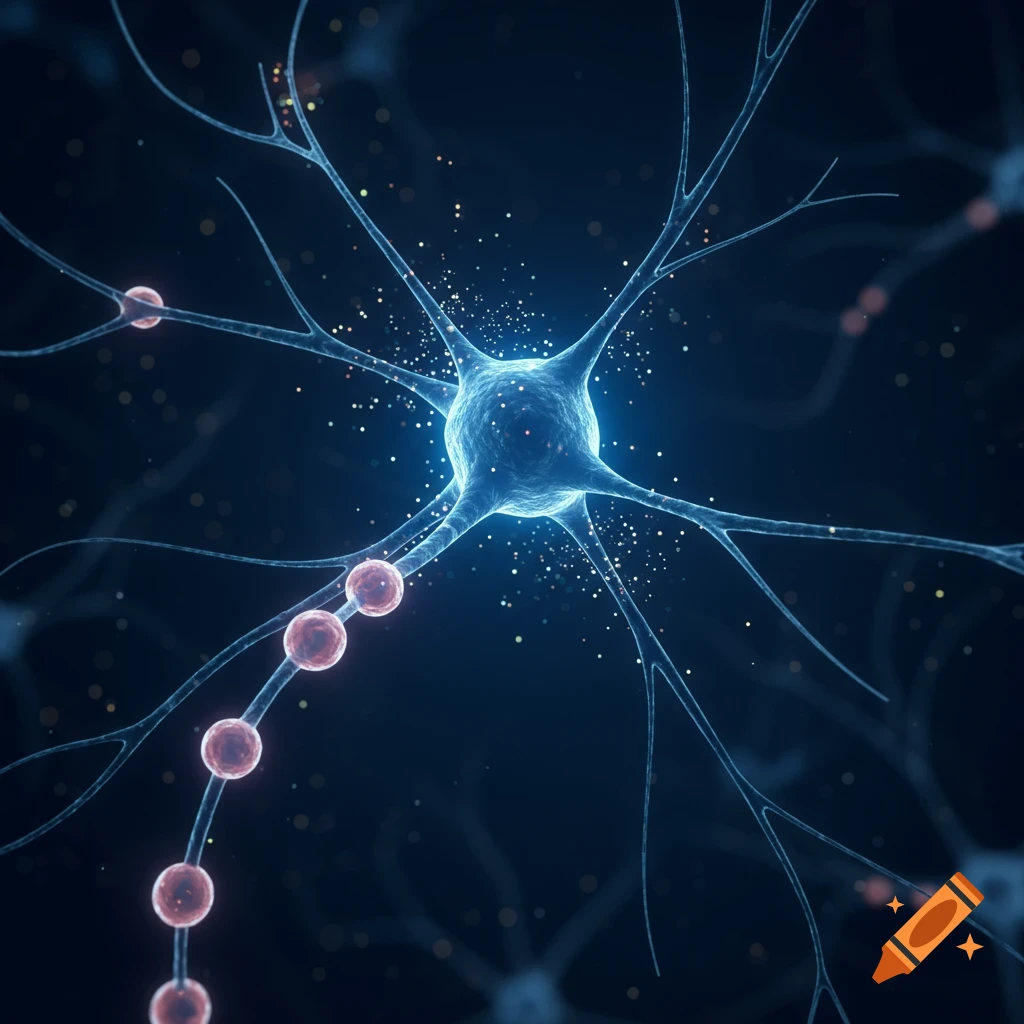
A detailed, stylized close-up rendering of a nerve cell with numerous branching dendrites and an axon, against a blurry blue and purple background.
Zoom-Aktion
A detailed 3D rendering of an animal cell, showing a large pink nucleus, red mitochondria, and other organelles within a translucent membrane.
Zoom-Aktion
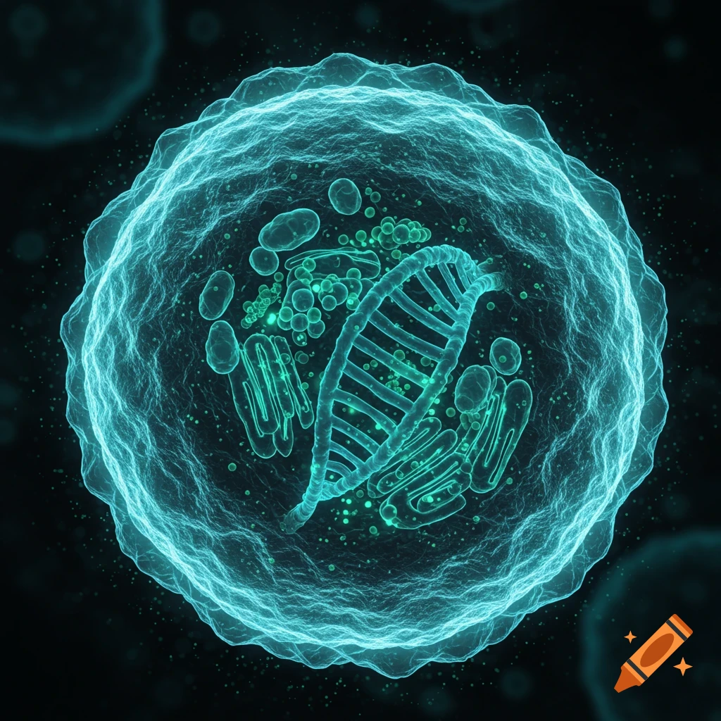
Stylized microscopic view of a translucent human cell with glowing, colorful organelles and a central helical structure.
Zoom-Aktion
A highly realistic microscopic view of adipose connective tissue with round adipocytes in a honeycomb pattern, stained pink and yellow.
Zoom-Aktion
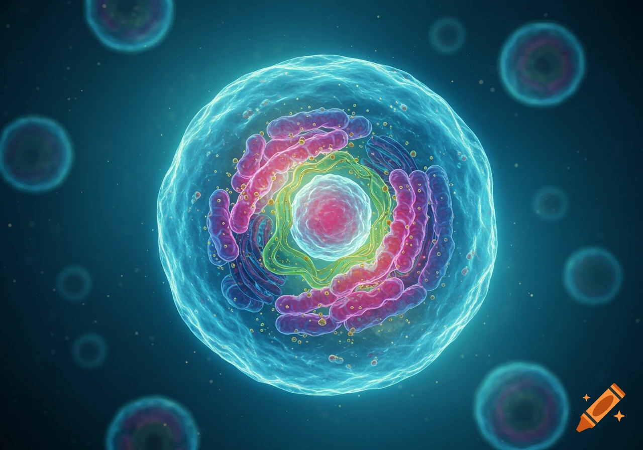
A vibrant, highly detailed cross-section of a eukaryotic cell, showcasing its intricate organelles and glowing nucleus in a blue-green environment.
Zoom-Aktion
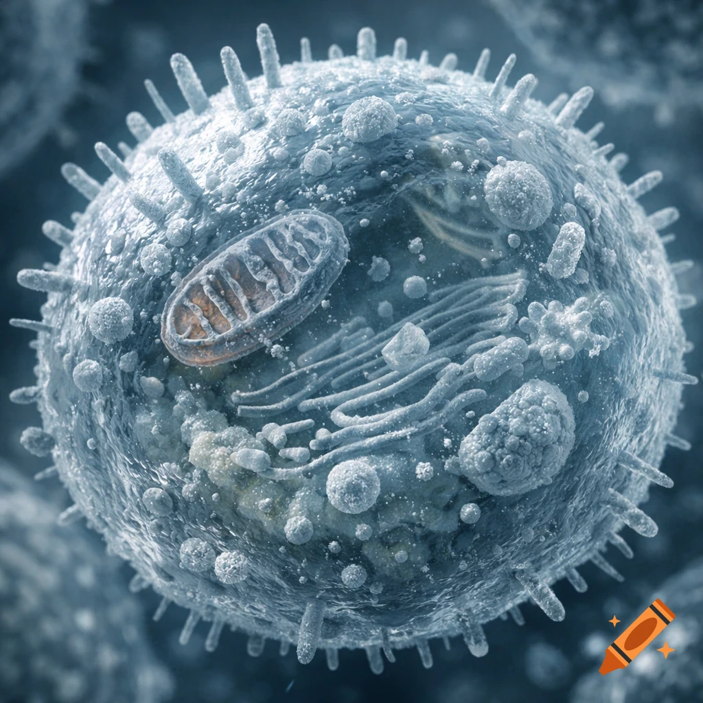
A detailed 3D rendering of an animal cell, showing internal organelles and a textured surface, in a scientific microscopy style.
Zoom-Aktion
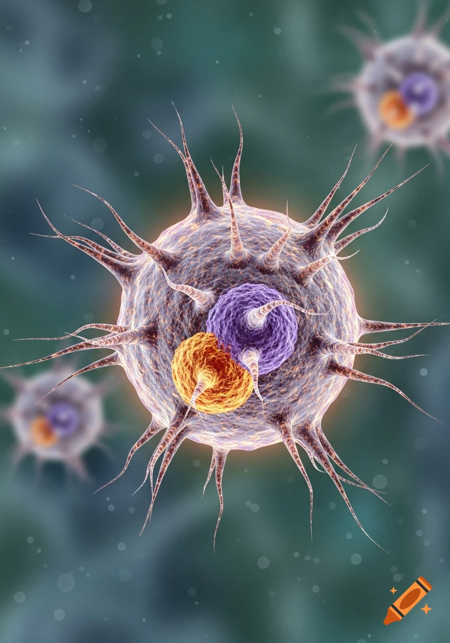
Close-up 3D rendering of a spiky, translucent cell with purple and orange internal components, against a blurry green background.
Zoom-Aktion
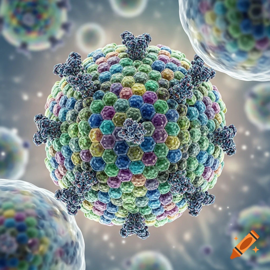
A detailed 3D illustration of a colorful virus particle, composed of hexagonal units, with spike proteins, against a blurred microscopic background.
Zoom-Aktion
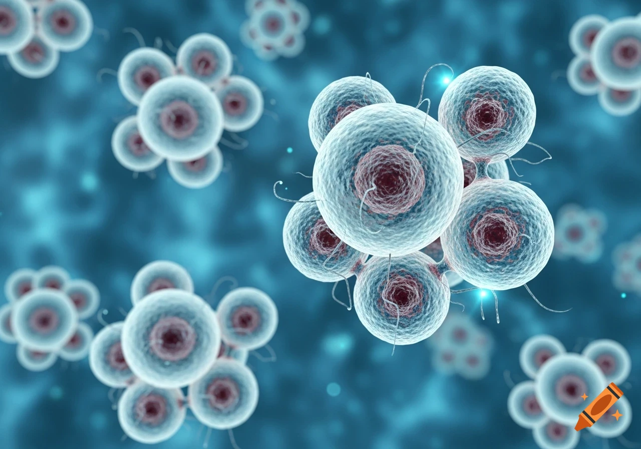
Close-up of numerous light blue, spherical cells with red centers and small filaments, against a blurry blue background.
Zoom-Aktion
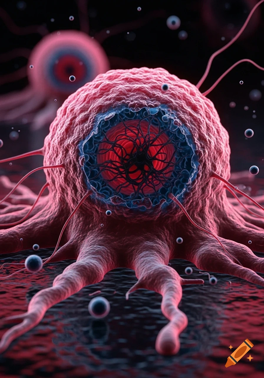
Close-up of a stylized red cancer cell with a dark blue and red core, tendrils, and floating particles on a dark background.
Zoom-Aktion
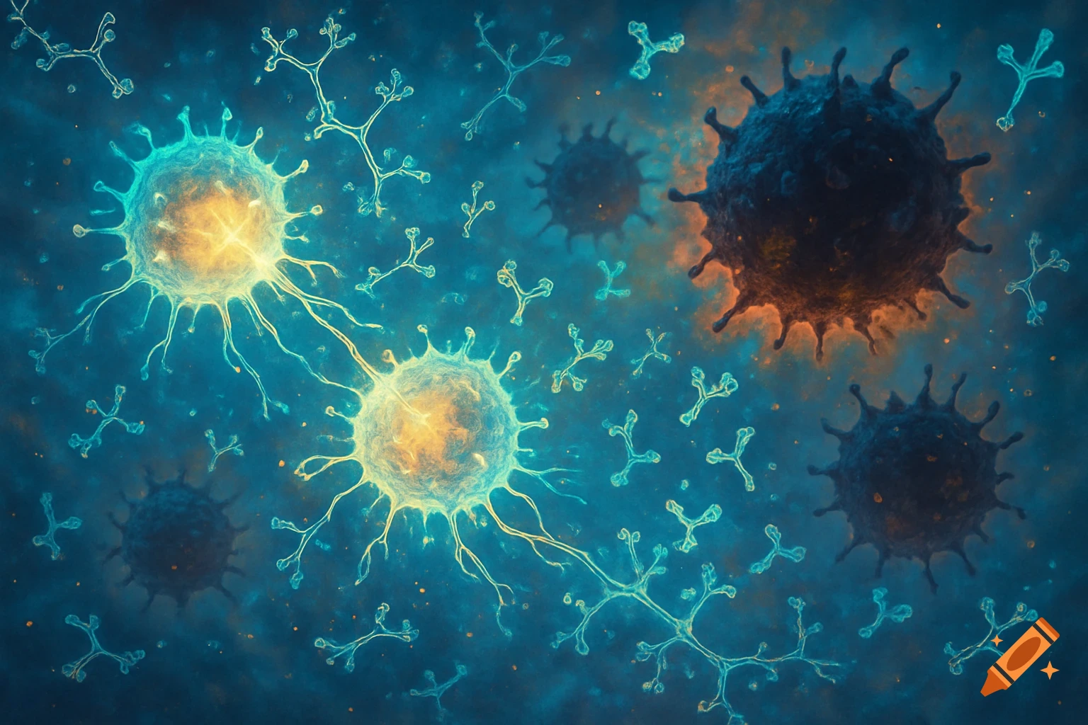
A microscopic illustration of glowing cells, dark spiky viruses, and light blue antibodies in a teal fluid.
Zoom-Aktion
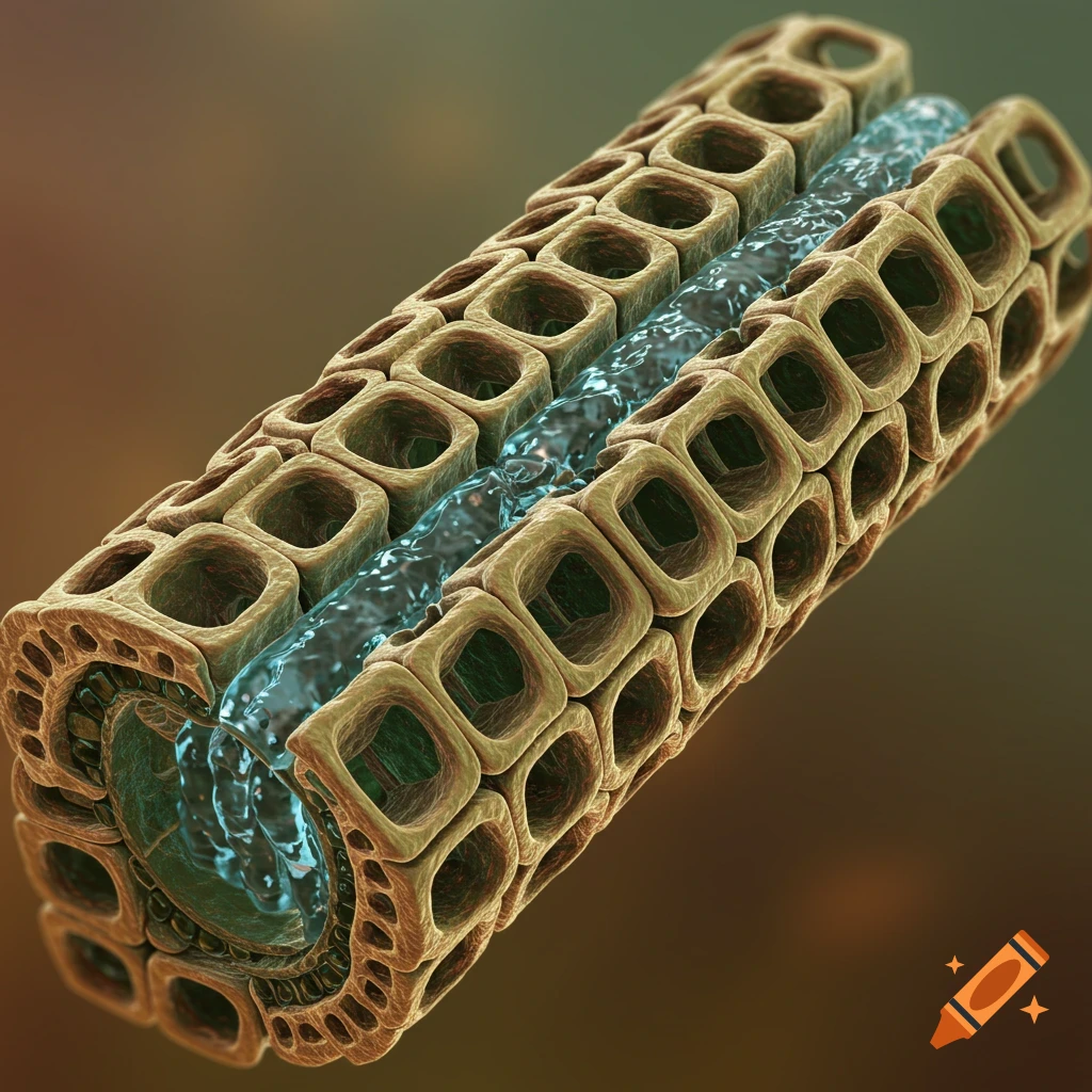
A detailed 3D cross-section of plant xylem, showing a central blue channel of water flowing through honeycomb-like cellular structures.
Zoom-Aktion
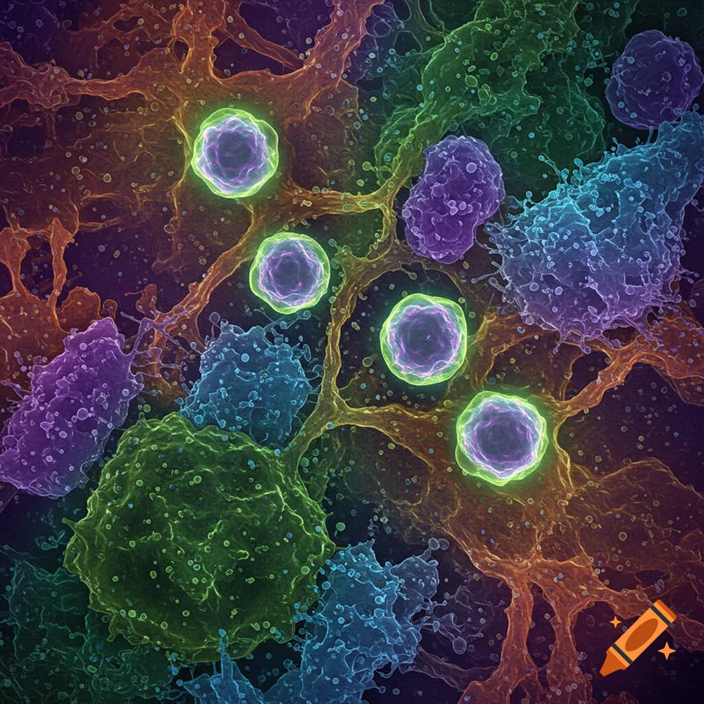
Vibrant microscopic image showing glowing green and purple cells interconnected within a complex orange and blue tissue network.
Zoom-Aktion
A 3D illustration of a biological nanopore in a lipid bilayer, surrounded by purple and blue spherical cells.
Zoom-Aktion
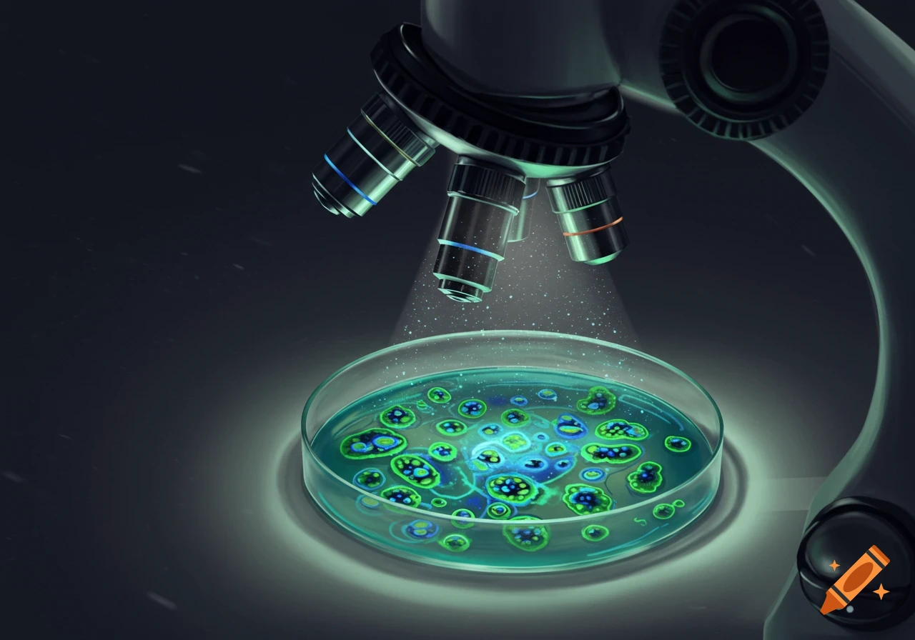
A close-up view of a microscope observing glowing green and blue microorganisms in a petri dish in a dark room.
Zoom-Aktion
A translucent single-cell organism with green and blue internal structures, surrounded by thin tendrils, floats in a dark teal liquid with smaller organisms.
Zoom-Aktion
A microscopic, heart-shaped cell or organism with a dark purple nucleus and radiating dendrite-like extensions, set against a dark purple background with faint molecular structures.
Zoom-Aktion
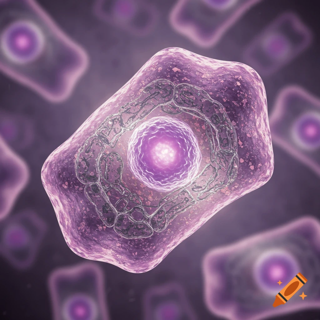
A vibrant purple, trapezoid-shaped cell with a central nucleus and intricate internal structures, viewed under a microscope.
Zoom-Aktion
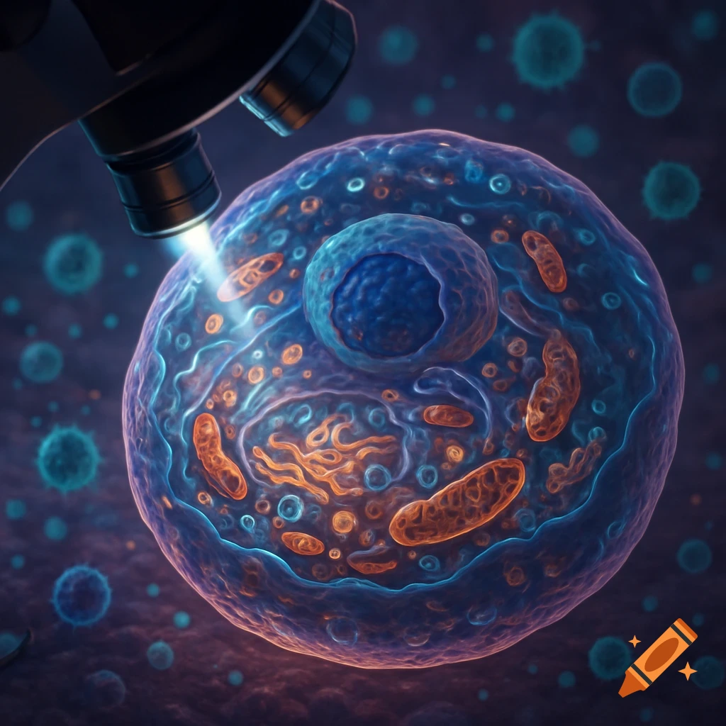
Detailed, stylized illustration of a cell under a microscope, illuminated by blue light, with internal organelles in orange and blue.
Zoom-Aktion
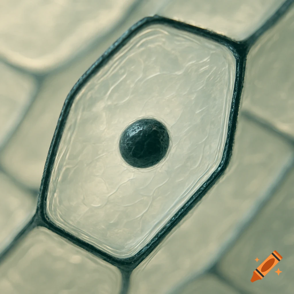
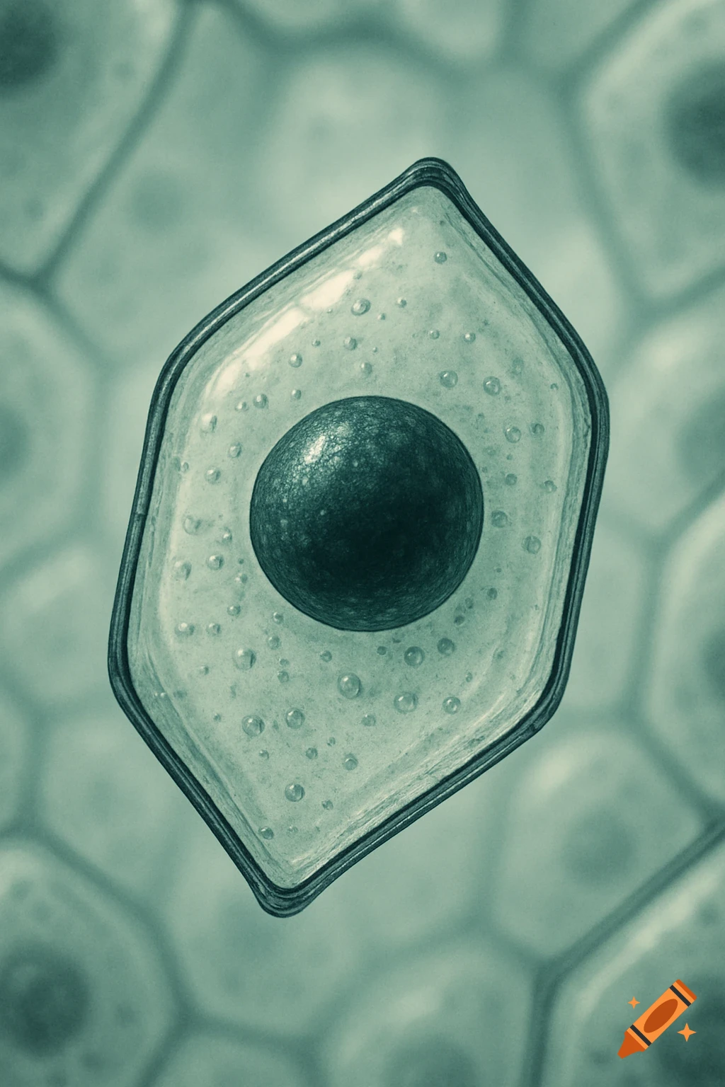
Close-up microscopic view of an elongated, six-sided onion cell with a dark nucleus within transparent cytoplasm.
Zoom-Aktion
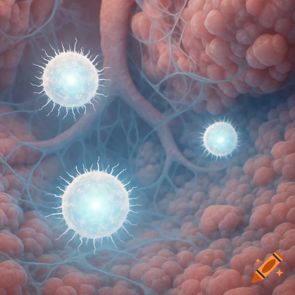
Microscopic view of three glowing white spiky cells against pink tissue and blue filaments.
Zoom-Aktion
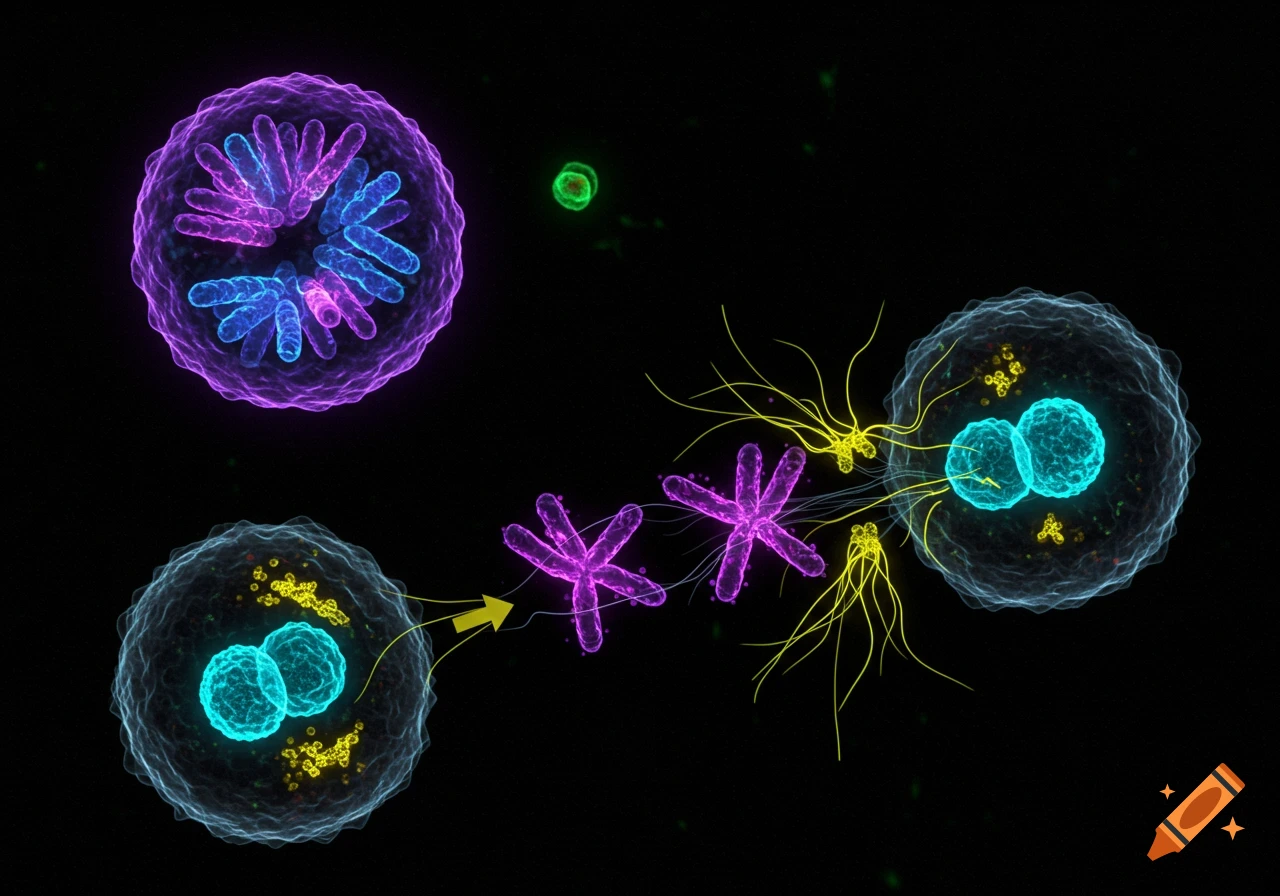
A microscopic view of animal cell mitosis with various fluorescently colored cells, chromosomes, and cellular components on a black background.
Zoom-Aktion
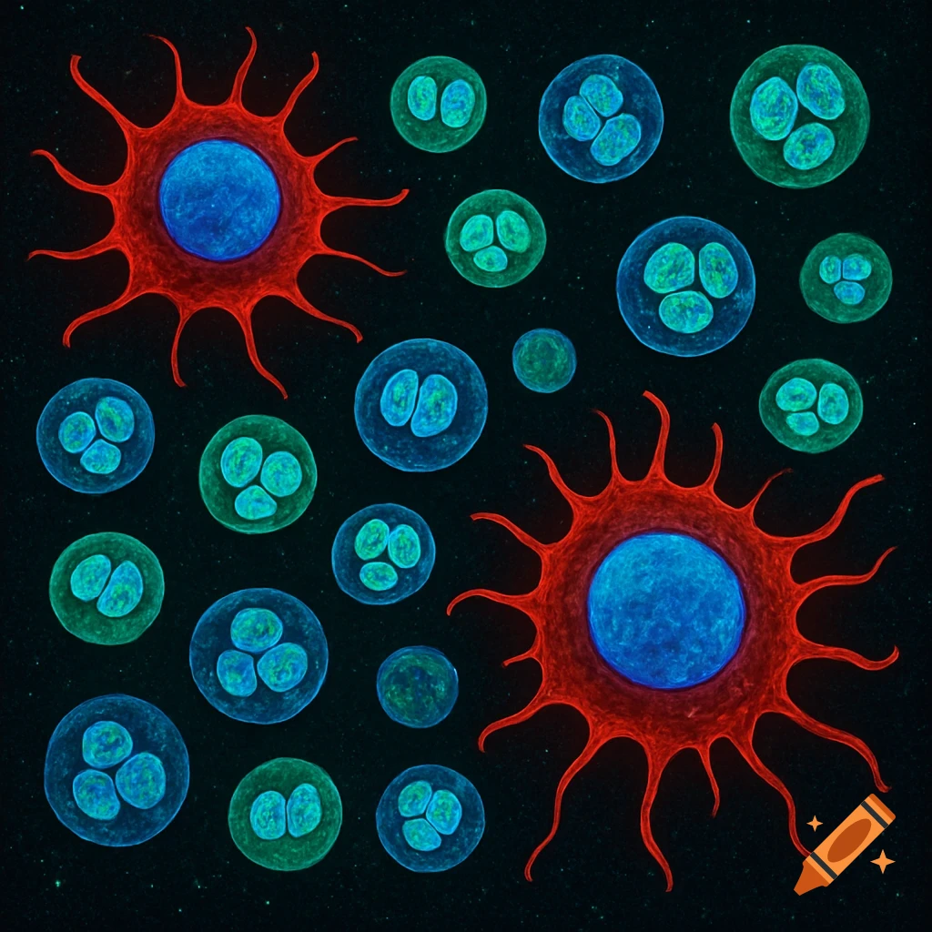
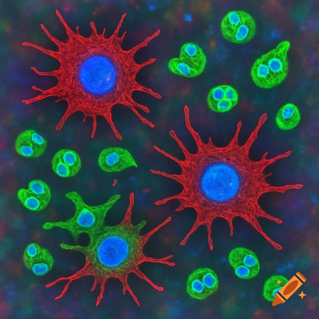
A colorful immunofluorescence image shows large red macrophages with blue nuclei and smaller green neutrophils with blue lobulated nuclei, against a dark, speckled background.
Zoom-Aktion
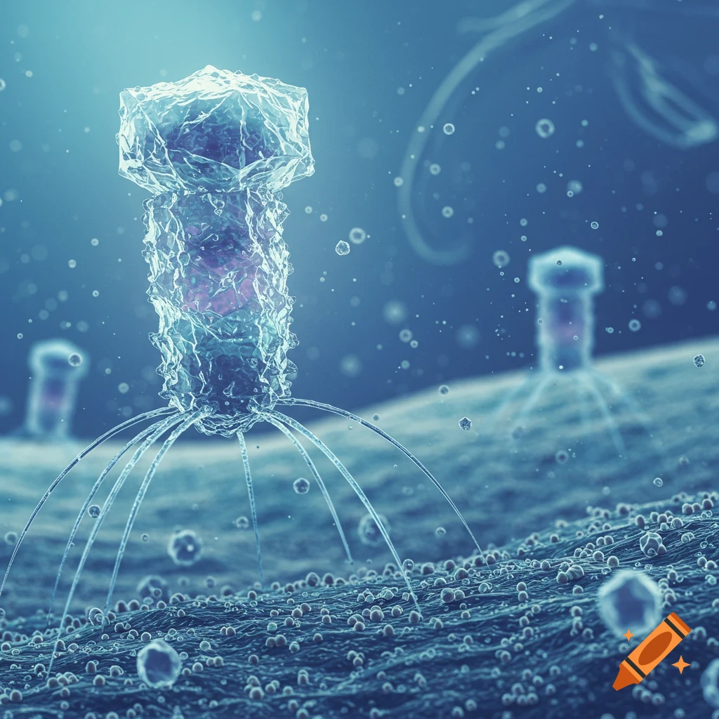
Detailed 3D render of a bacteriophage with an angular head and leg-like structures on a textured blue surface, surrounded by other microscopic particles.
Zoom-Aktion
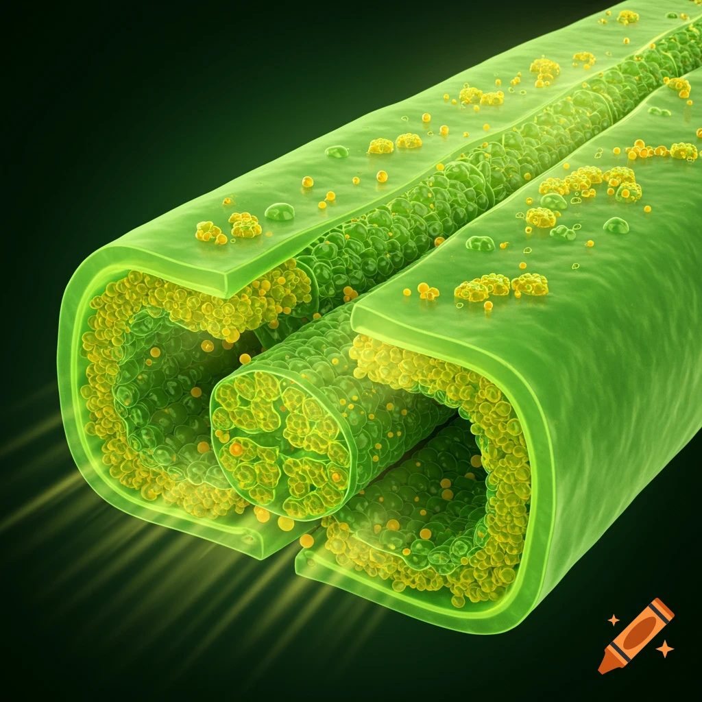
A detailed 3D scientific illustration of a chloroplast, showing a cross-section of its internal green and yellow structures.
Zoom-Aktion
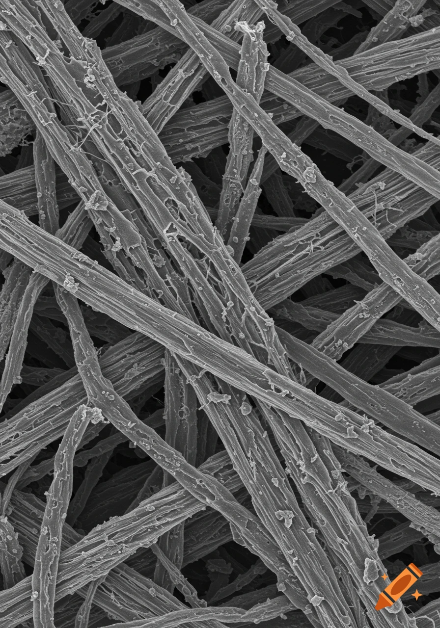
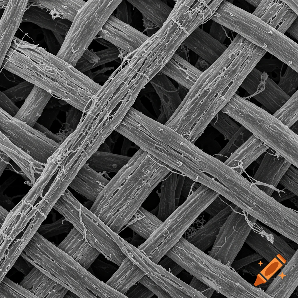
A grayscale scanning electron microscope image of interwoven aramid fibers forming a mesh.
Zoom-Aktion
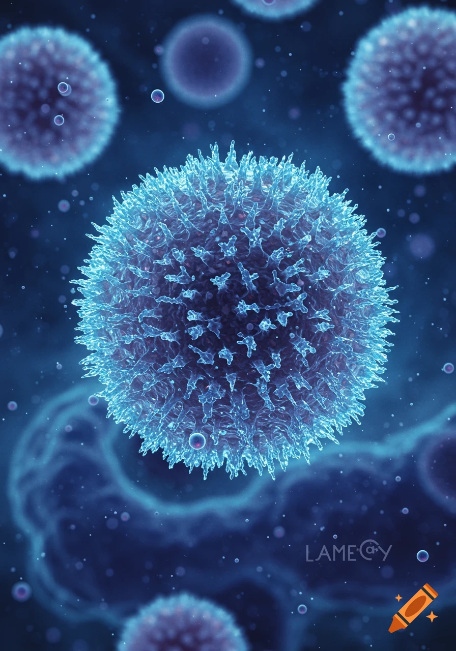
A vibrant blue microscopic cell, resembling a virus or lymphocyte, with spike-like projections in a dark blue cellular environment.
Zoom-Aktion
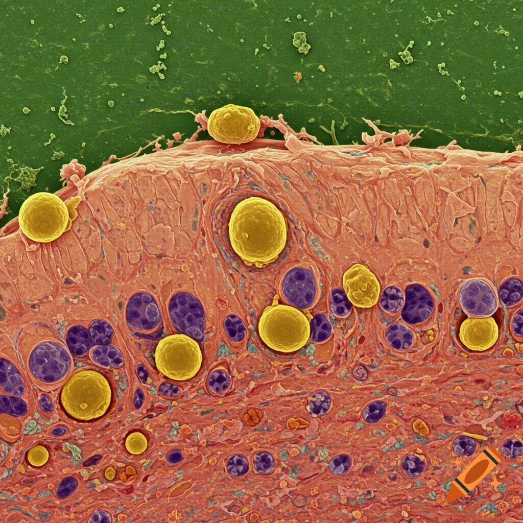
A colorful microscopic image showing yellow and purple cells within a reddish-pink tissue matrix, characteristic of cartilage.
Zoom-Aktion
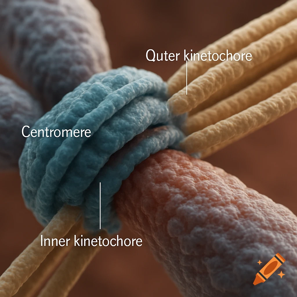
A photorealistic close-up view of a centromere (blue) with inner (blue) and outer (orange) kinetochores, and attached microtubules (yellow), labeled.
Zoom-Aktion
Microscopic view of pink epithelial cells with purple nuclei, surrounded by smaller round cells.
Zoom-Aktion
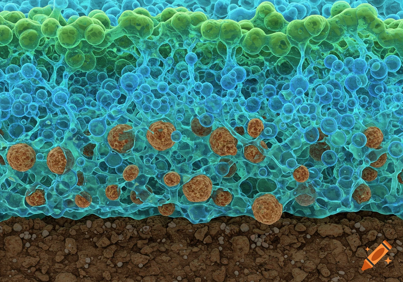
Microscopic view of green and blue gel-like substance binding brown soil particles.
Zoom-Aktion
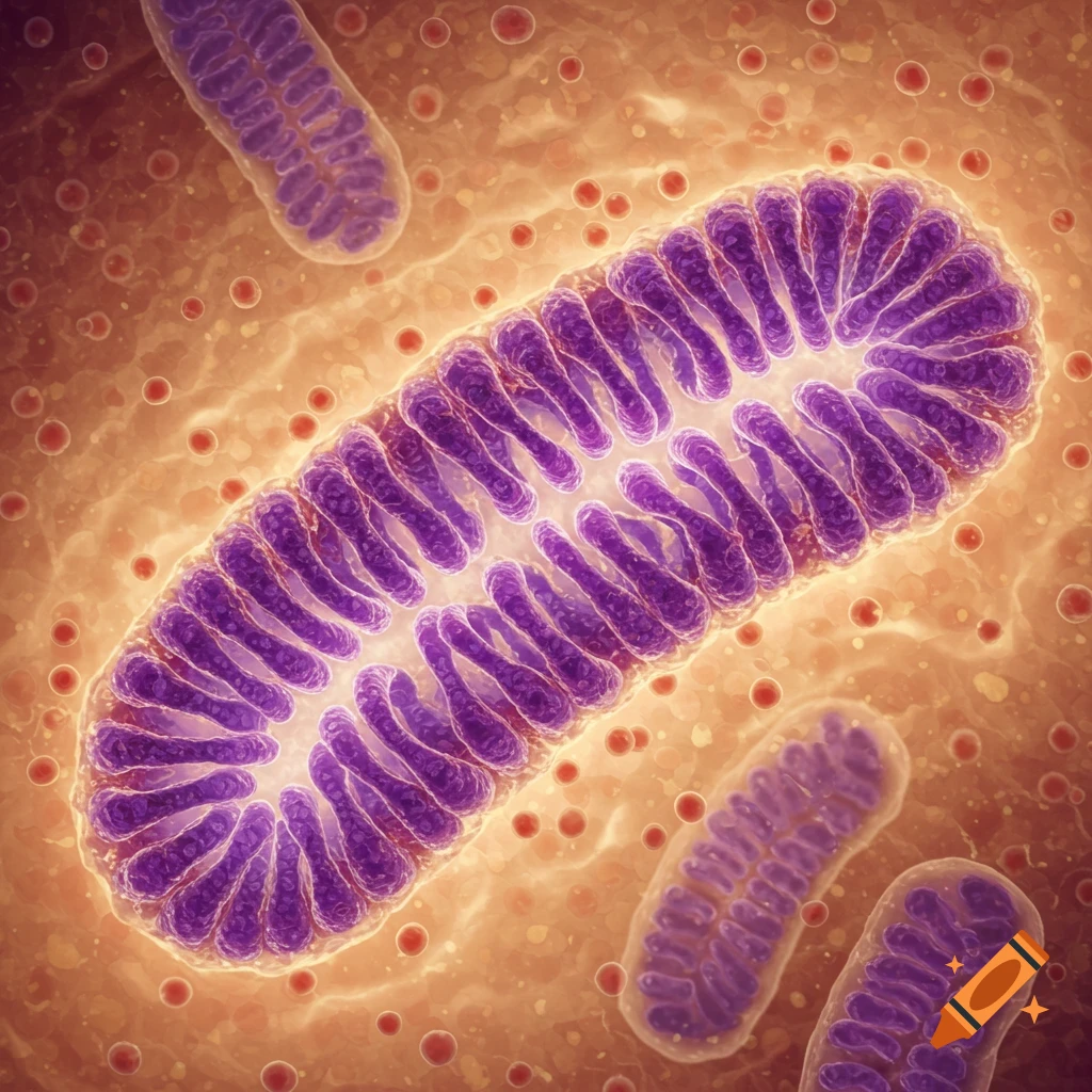
Close-up microscopic view of purple mitochondria structures inside a cell, glowing with light.
Zoom-Aktion
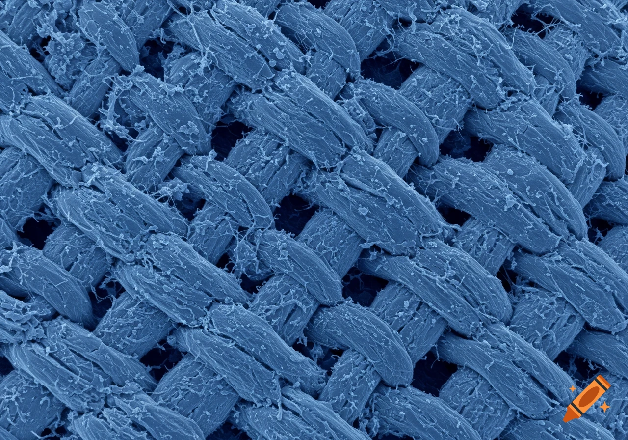
Close-up view of blue fabric weave under an electron microscope.
Zoom-Aktion
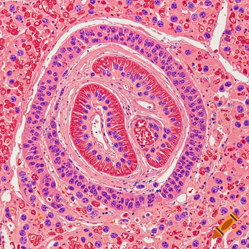
Close-up microscopic view of biological tissue, showing cellular structure in pink and purple.
Zoom-Aktion
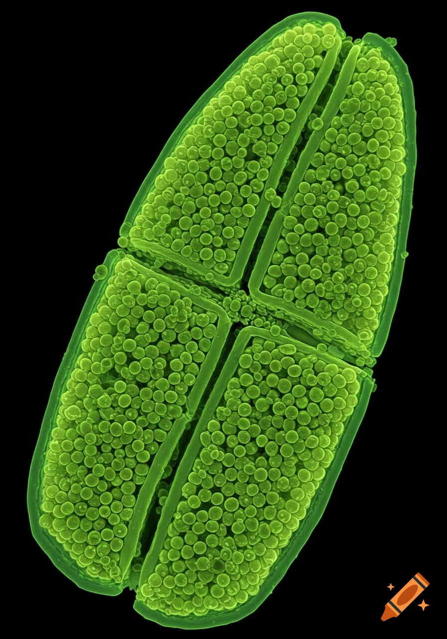
Close-up microscopic view of a green segmented structure filled with spherical details on a black background
Zoom-Aktion
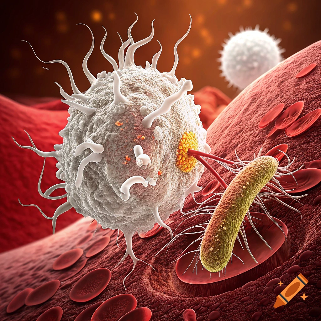
Microscopic view of a white blood cell attacking a bacterium in a blood vessel.
Zoom-Aktion
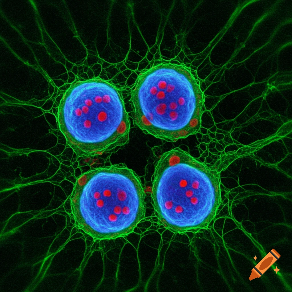
Four stylized blue cell-like shapes with red dots surrounded by green network on black background, microscopic view.
Zoom-Aktion
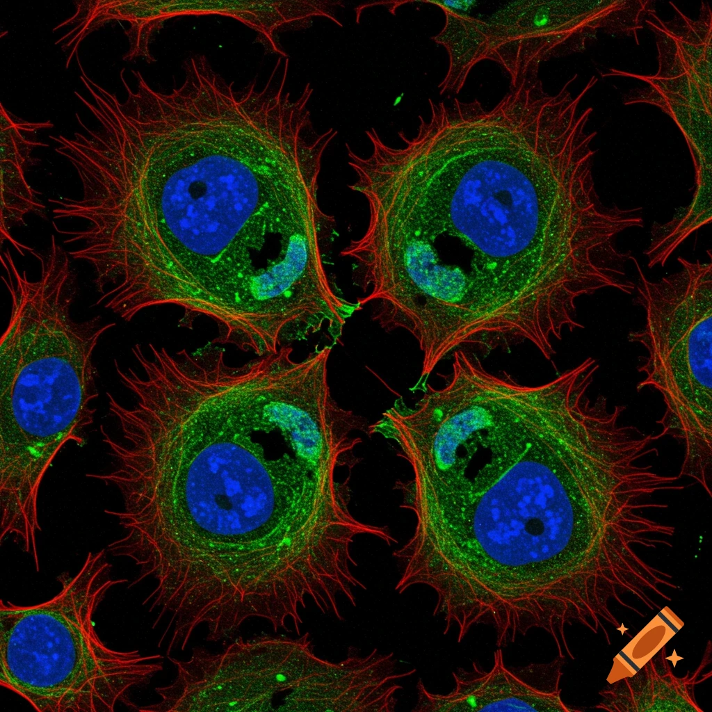
Fluorescence microscopy image of cells with green actin, red microtubules, and blue nuclei on a black background.
Zoom-Aktion
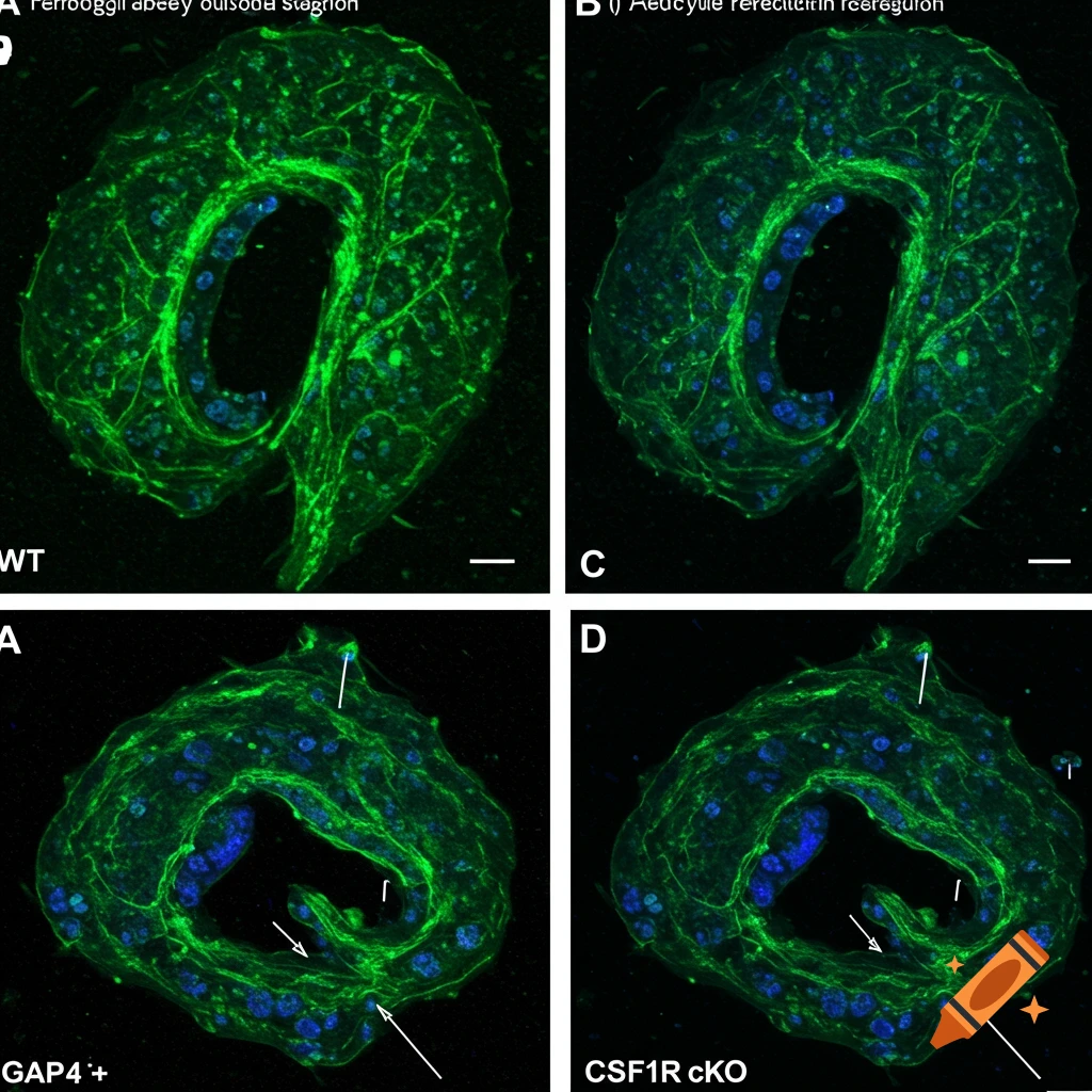
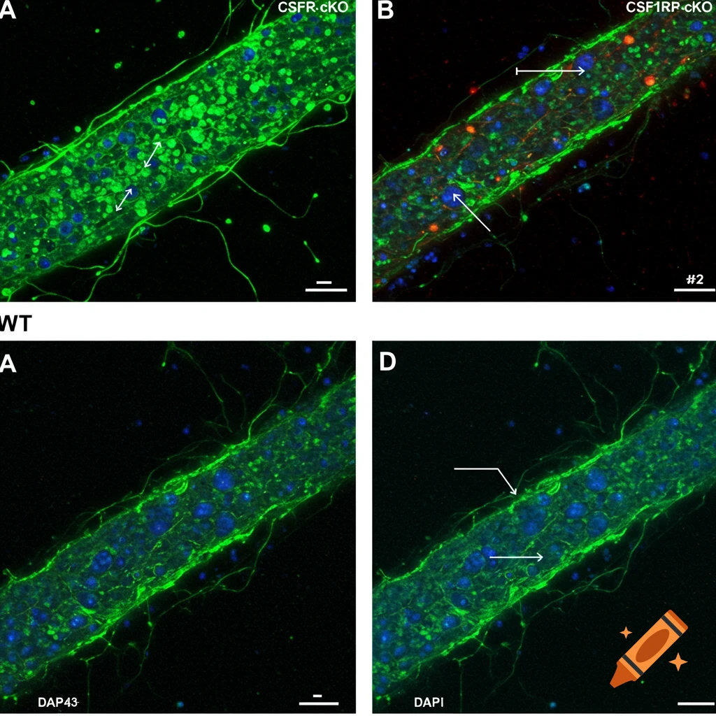
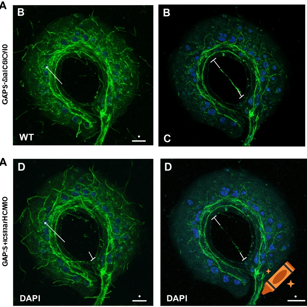
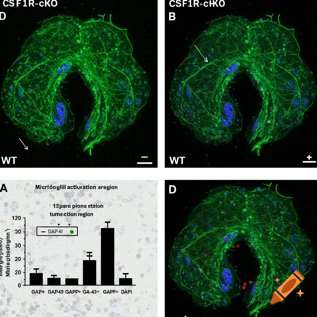
Immunofluorescence images of GAP-43 staining showing axonal regrowth post-tumor resection
Zoom-Aktion
Microscopic view of vibrant bacterial biofilm with diverse morphologies
Zoom-Aktion
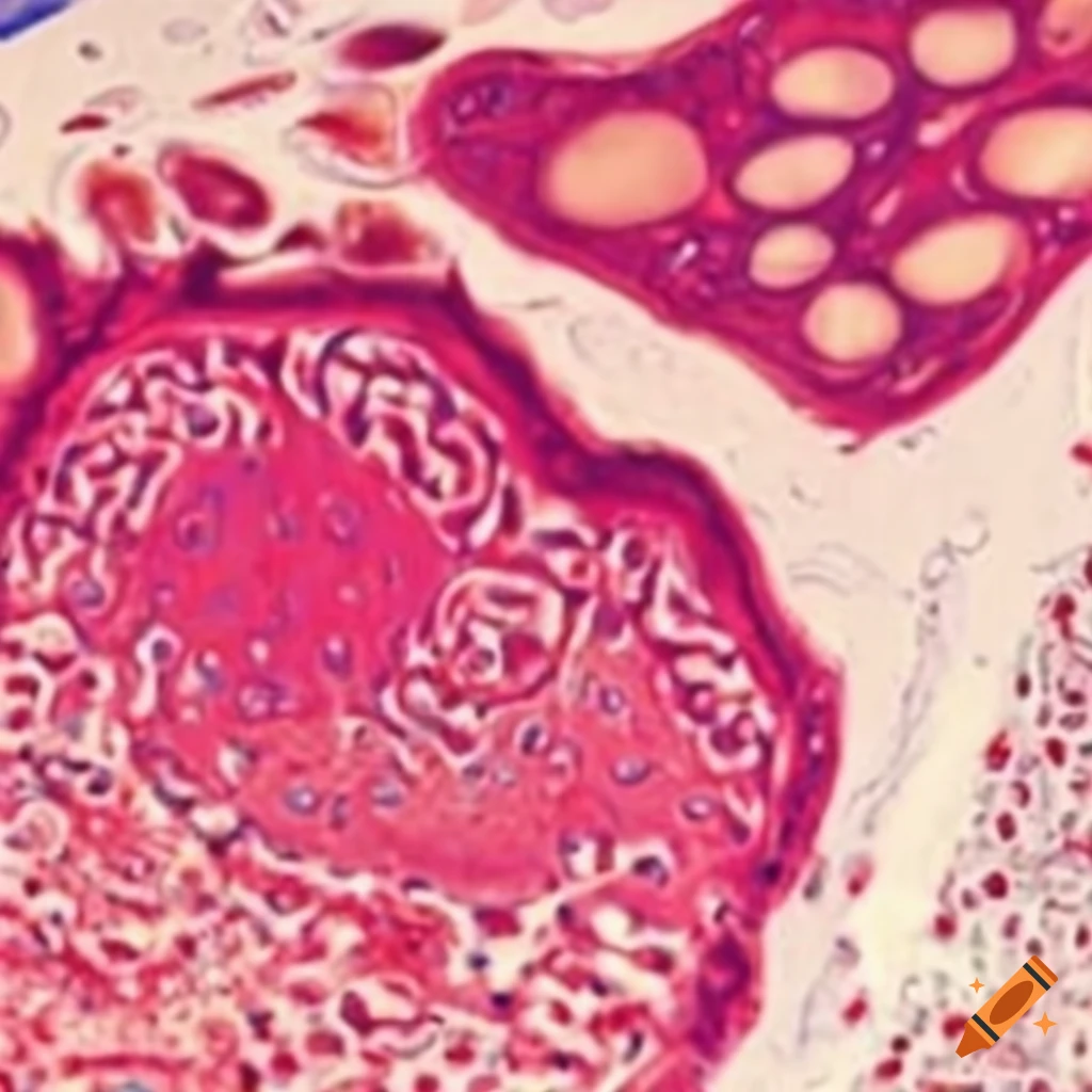
Microscopic view of stained tissue sample with varying cell sizes and shapes
Zoom-Aktion
Bromine-yeast cell viewed under microscope
Zoom-Aktion
Detailed microscopic image showcasing various human cells
Zoom-Aktion
Cell structure under a microscope
Zoom-Aktion
Inclusion microstructure
Zoom-Aktion
Genetics and electron microscope in high definition
Zoom-Aktion
Genetics and electron microscope in high definition
Zoom-Aktion
Colorful super resolution microscopy image of a pancreas
Zoom-Aktion
Surrealism under a microscope with fine detail
Zoom-Aktion
Detailed visualization of a single cell bacterium
Zoom-Aktion
3D representation of fluorescent gene transcripts in tissue slide
Zoom-Aktion
High magnification of a tungsten filament in an electron microscope
Zoom-Aktion
Magnified intricate fungal structures in a culture petri dish
Zoom-Aktion
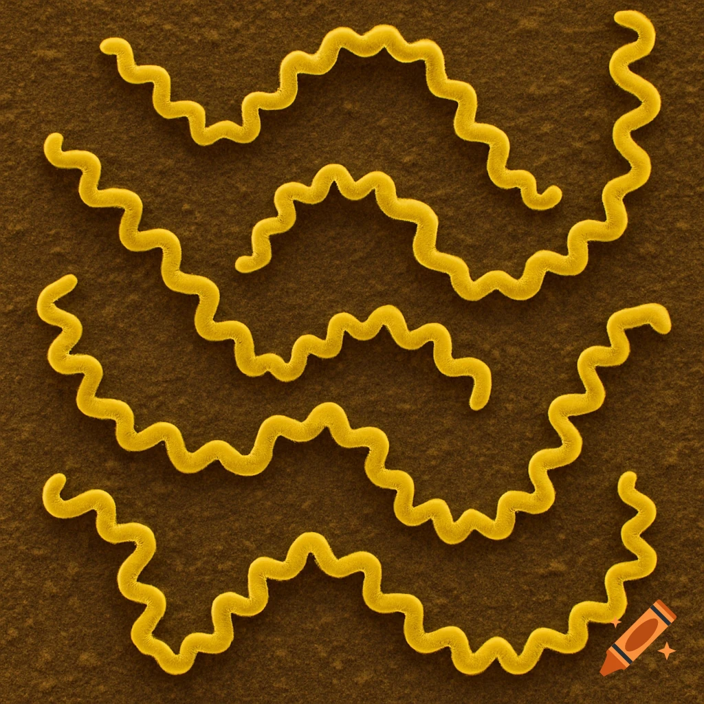
Microscopic view of long curly bacteria
Zoom-Aktion
Workbook cover on histology and embryology with microscope and scientist
Zoom-Aktion
High magnification of translucent membrane with histological stains
Zoom-Aktion
Cobalt oxide SEM super detailed image on black background
Zoom-Aktion
Cell
Zoom-Aktion
Microscopic view
Zoom-Aktion
Microscopic view
Zoom-Aktion
Green and orange logo for microscopy expertise and training on black background
Zoom-Aktion
Genetics and electron microscopy exploring the intricate tapestry of life at a hyperdetailed level with 3D renders
Zoom-Aktion
Genetics and electron microscopy exploring the intricate tapestry of life at a hyperdetailed level with 3D renders
Zoom-Aktion
Antibody staining
Zoom-Aktion
Microscopic view of a brain
Zoom-Aktion
electron microscopy of yeast cells
Zoom-Aktion
seamless pattern of human lung cells under a microscope
Zoom-Aktion
scientist examining viruses through a microscope
Zoom-Aktion
close-up of a mitochondria
Zoom-Aktion
close-up of metal mesh separating gas flow
Zoom-Aktion
illustration of the Little Prince using an electron microscope
Zoom-Aktion
microscopic view of gold titanium oxide particles
Zoom-Aktion
fractal fungal growth inspired by Haeckel
Zoom-Aktion
fractal fungal growth inspired by Haeckel
Zoom-Aktion
cells colliding in a microscopic view
Zoom-Aktion
microscopic view of a eukaryotic cell with nucleus and lipid bilayer
Zoom-Aktion
cells making protein in a biological process
Zoom-Aktion
microscopic view of a crystal diamond texture
Zoom-Aktion
fluorescent microscopy image of bacteria
Zoom-Aktion
microscope image
Zoom-Aktion
logo design of Ai meets microscopy
Zoom-Aktion
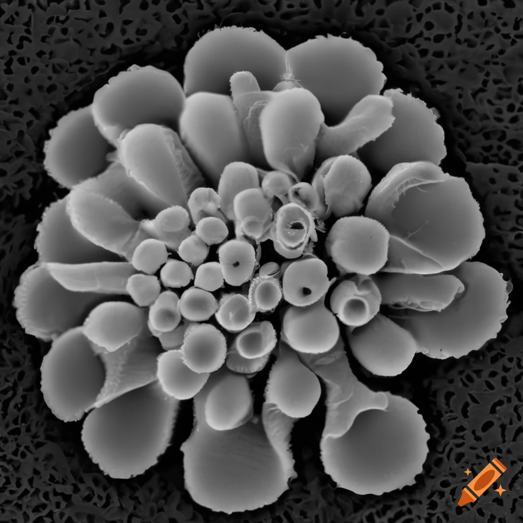
black and white scanning electron microscopy of a goat willow flower
Zoom-Aktion
black and white microscopy of a rhododendron flower
Zoom-Aktion
black and white microscopy of a buddleja flower
Zoom-Aktion
black and white image of a snowflake under electron microscopy
Zoom-Aktion
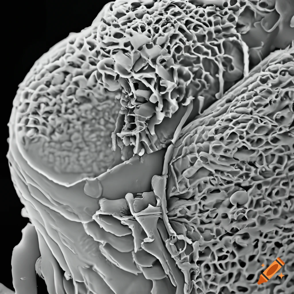
scanning electron microscopy of lichen cells
Zoom-Aktion
black and white SEM image of borage
Zoom-Aktion
black and white microscopy image of lichen
Zoom-Aktion
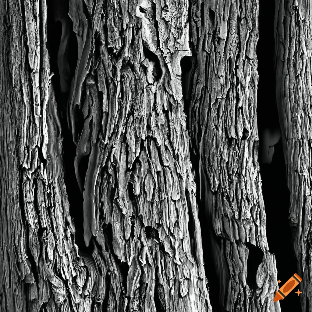
black and white microscopic image of tree bark
Zoom-Aktion
black and white microscopic image of flower pollen
Zoom-Aktion
SEM image of a feather
Zoom-Aktion
microscopic image of a cell
Zoom-Aktion
view of human gallbladder cells under the microscope
Zoom-Aktion
microscopy of human uterine cells
Zoom-Aktion
close-up of pancreatic cells under a microscope
Zoom-Aktion
artistic depiction of endoplasmic reticulum-mitochondria contact sites
Zoom-Aktion
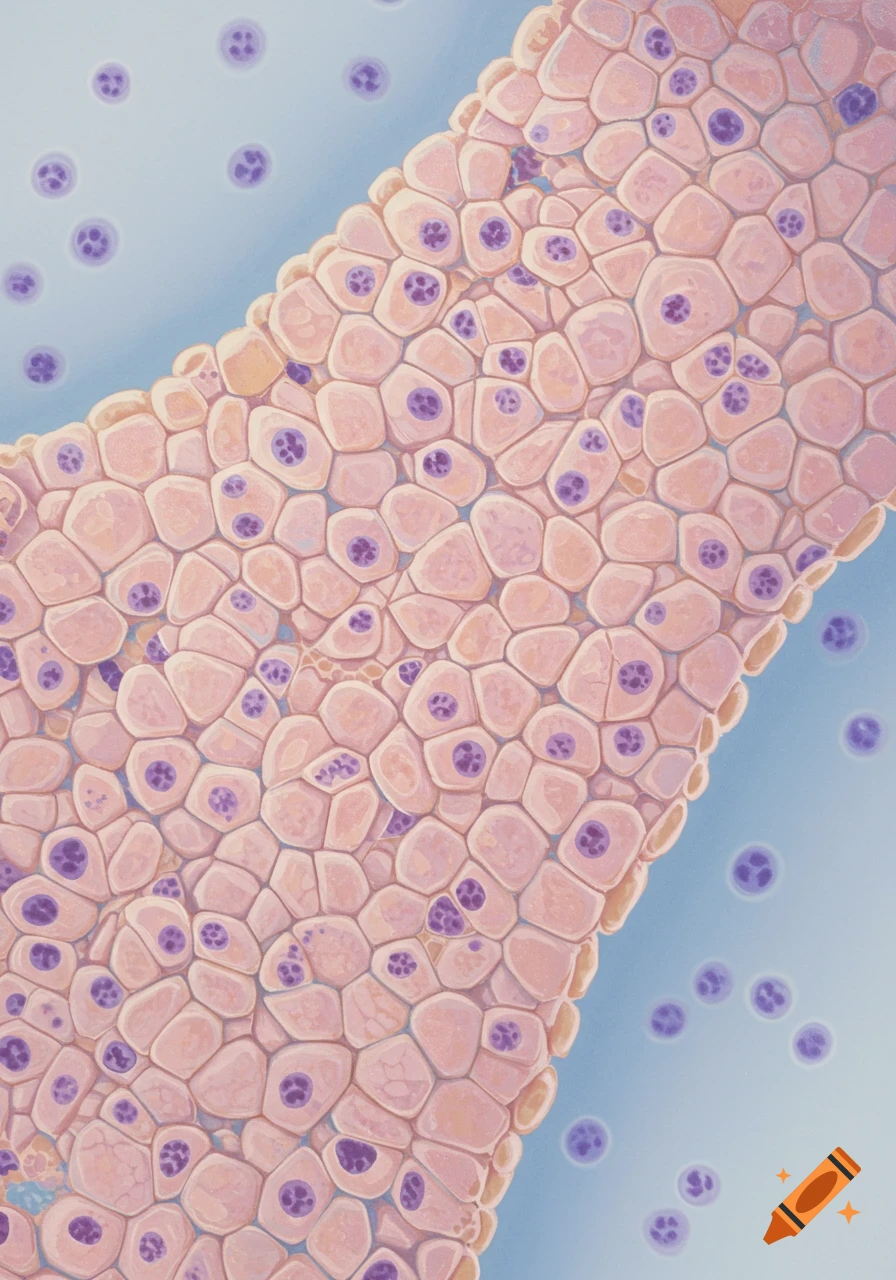
microscopic view of human oral mucosa cells
Zoom-Aktion
microscopic view of human lung cells
Zoom-Aktion
detailed depiction of biological microorganisms
Zoom-Aktion
microscopic view of a human bone cell
Zoom-Aktion
microscopic view of human muscle cells
Zoom-Aktion
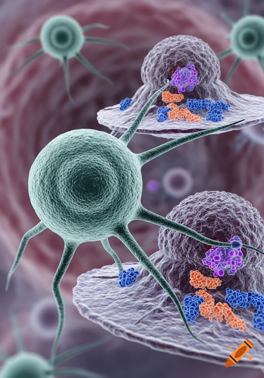
image of a vibrant Vitamin B12 cell
Zoom-Aktion
microscopic image of a bumpy animal cell
Zoom-Aktion
microscopic view of blood cells
Zoom-Aktion
Photorealistic microscopic view of blue and green cells with glowing data points and digital connections on a blue-gray background.
Zoom-Aktion
A vibrant fluorescent micrograph showing green and blue neural tissue with intricate connections on a black background.
Zoom-Aktion
A microscopic view of a Y-shaped antibody in blue and green, surrounded by red blood cells and other organic structures.
Zoom-Aktion
A detailed, translucent biological cell showing a large nucleus, mitochondria, and other organelles against a blurry lab background.
Zoom-Aktion
A 3D rendering of a purple coronavirus particle with red spike proteins on a blurry gray background.
Zoom-Aktion
A stylized immunofluorescence image of a cell with a green actin network, red Golgi apparatus, and blue nucleus on a black background.
Zoom-Aktion
A detailed 3D render of a rod-shaped rabies virus particle, showing an internal helix and surface glycoproteins, against a blue background.
Zoom-Aktion
A well-lit science laboratory with a microscope, various surgical tools, petri dishes with colorful cultures, and vials on a metal counter.
Zoom-Aktion
A detailed microscopic rendering of a vibrant purple and blue cell or virus with surface receptors and an intricate internal network against a dark background.
Zoom-Aktion
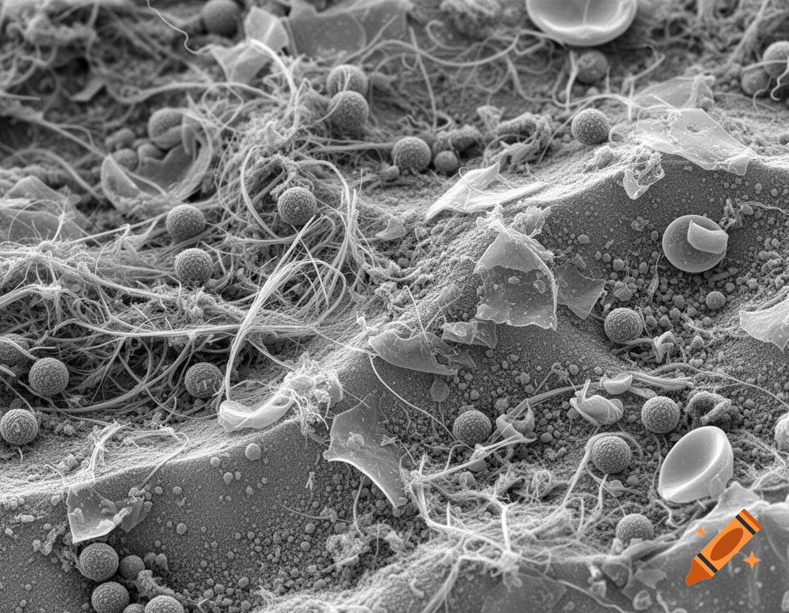
A detailed black and white electron microscope image of house dust, showing various fibers, spherical spores, and irregular particles.
Zoom-Aktion
A microscopic view of cells with green and blue chromosome-like structures connected by delicate filaments, seen through a magnifying glass.
Zoom-Aktion
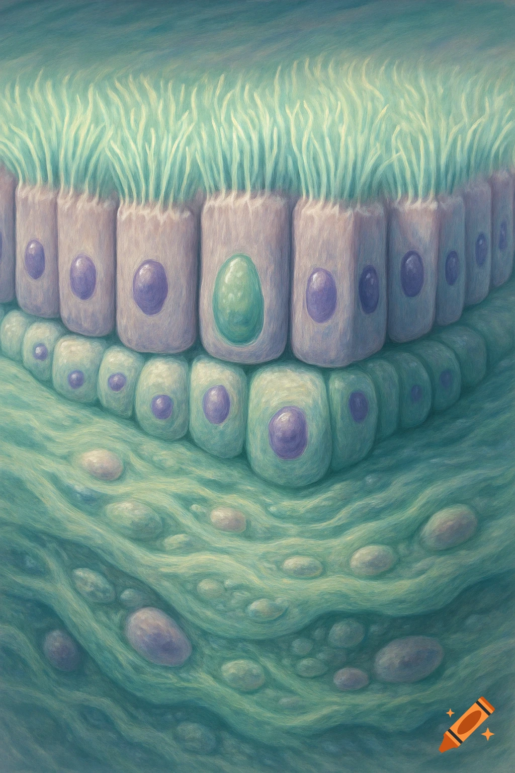
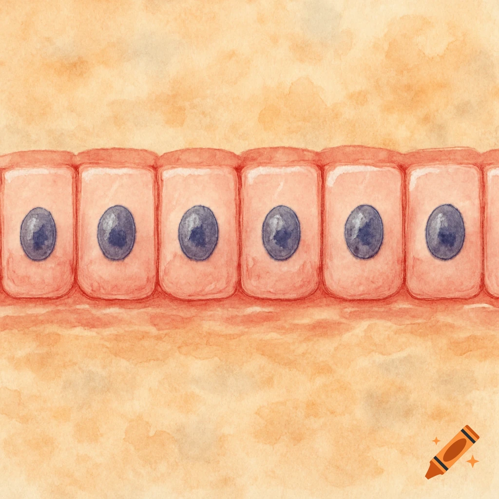
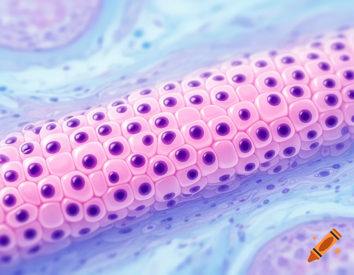
Close-up microscopic view of pink cuboidal cells with dark purple centers arranged in rows, against a blue and purple background.
Zoom-Aktion
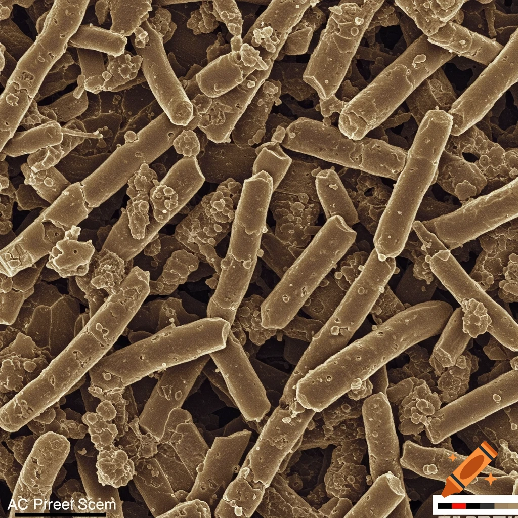
Microscopic view of blue rod-shaped bacteria among white and dark interwoven fibers against a hazy grey background.
Zoom-Aktion
Close-up microscopic view of green plant cells, showing polygonal cell walls and numerous chloroplasts within.
Zoom-Aktion
Vibrant illustration of a biological cell with organelles, glowing nucleus, and text 'Cell Biology & Microscopy' on a blue background.
Zoom-Aktion
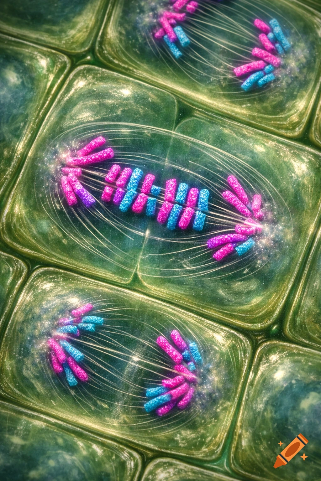
Microscopic view of plant cells undergoing mitosis, with colorful pink and blue chromosomes dividing inside green rectangular cells.
Zoom-Aktion
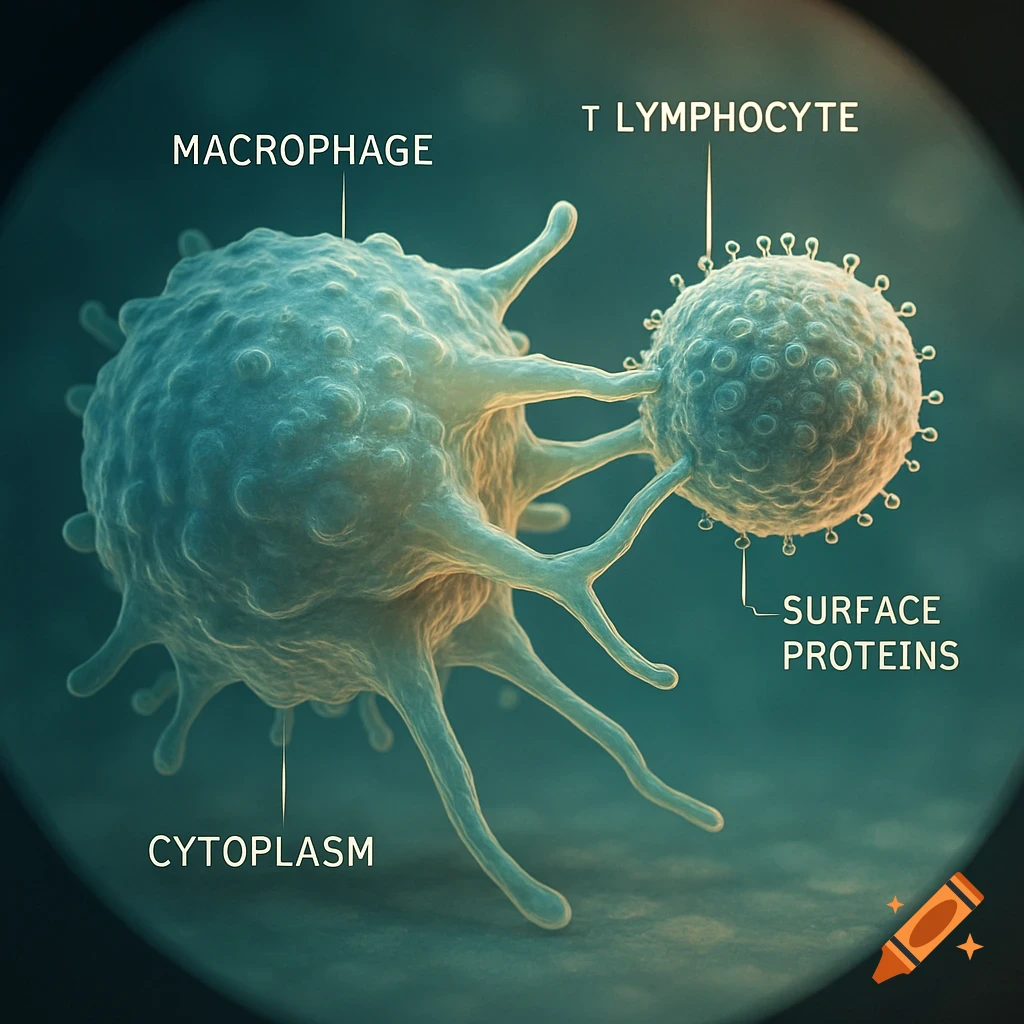
Microscopic scientific diagram of a macrophage with labeled cytoplasm interacting with a T lymphocyte and its surface proteins.
Zoom-Aktion
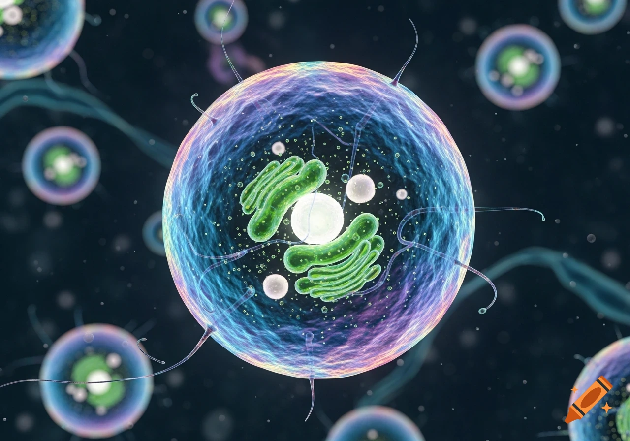
Close-up of a glowing iridescent cell with green internal structures and thin tendrils, against a dark background with blurred cells.
Zoom-Aktion
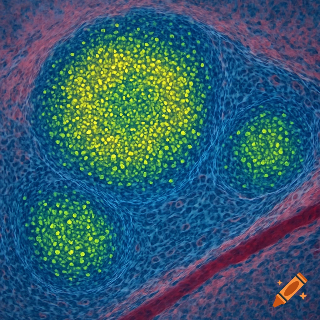
Microscopic view of three bright yellow-green cell clusters within blue and red biological tissue.
Zoom-Aktion
A small mouse-like mammal on a water lily leaf being observed under a detailed microscope with golden light trails.
Zoom-Aktion
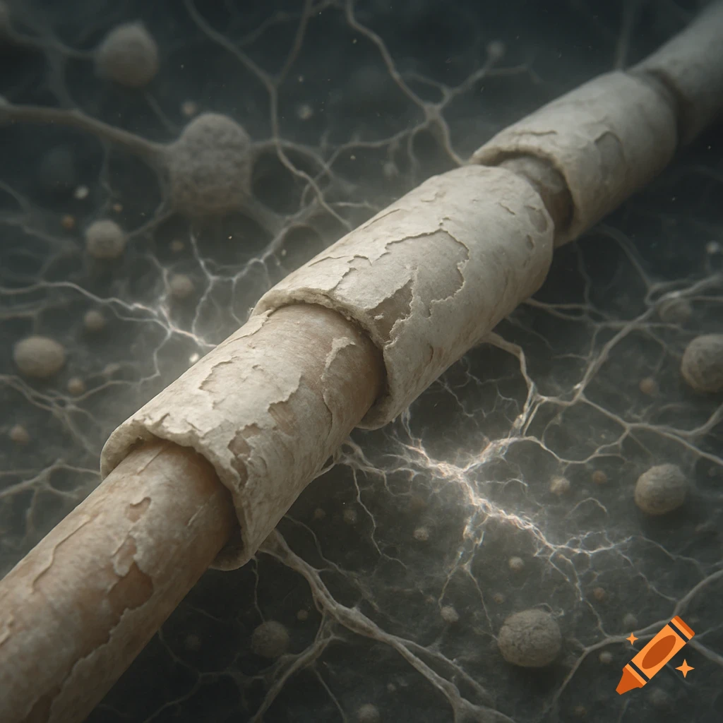
Microscopic view of a nerve fiber with a damaged, peeling myelin sheath and surrounding cellular network.
Zoom-Aktion
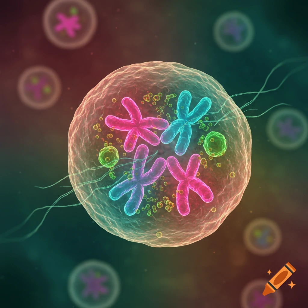
Vibrant stylized illustration of a cell undergoing division, with colorful X-shaped chromosomes and cellular structures.
Zoom-Aktion
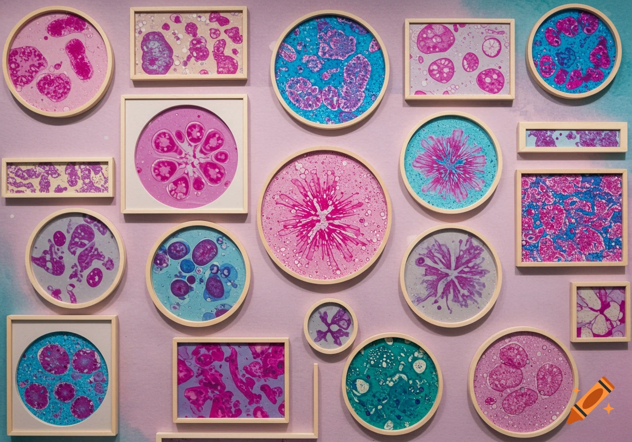
A wall covered with various framed abstract artworks, some round and some rectangular, featuring colorful microscopic patterns in pinks, blues, and purples.
Zoom-Aktion
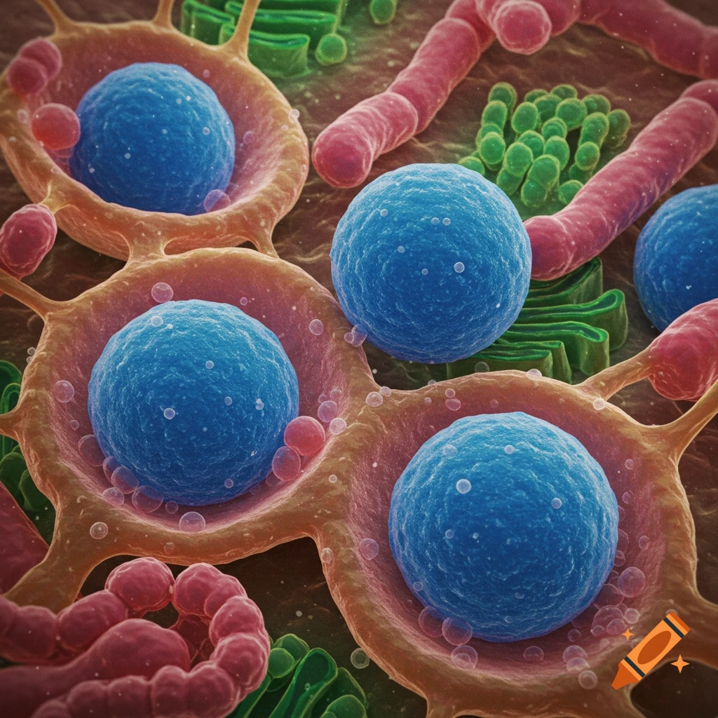
Microscopic view of blue spheres resembling lysosomes within cellular structures, alongside red and green tubular organelles.
Zoom-Aktion
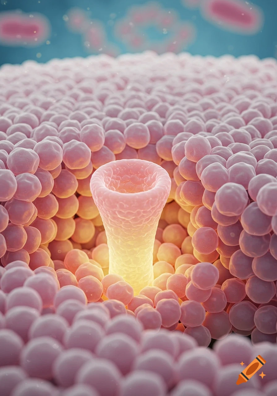
A detailed 3D rendering of a biological nanopore glowing within a pink lipid bilayer against a blue background.
Zoom-Aktion
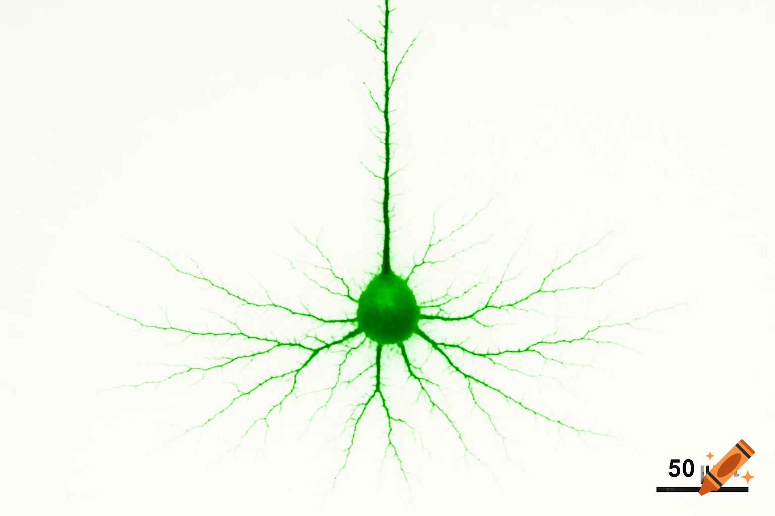
A vibrant green pyramidal neuron with extensive branching dendrites and an apical dendrite, against a white background, with a 50 5m scale bar.
Zoom-Aktion
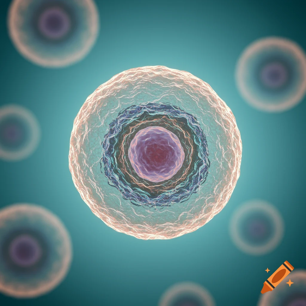
A translucent biological cell with a prominent purple nucleus centered against a blue background, depicting a microscopic view.
Zoom-Aktion
Close-up microscopic view of a cell with a purple nucleus, green inner layer, and blue outer layer.
Zoom-Aktion
Microscopic view of a teal-blue cell with a central nucleus, cytoplasm, and outer membrane.
Zoom-Aktion
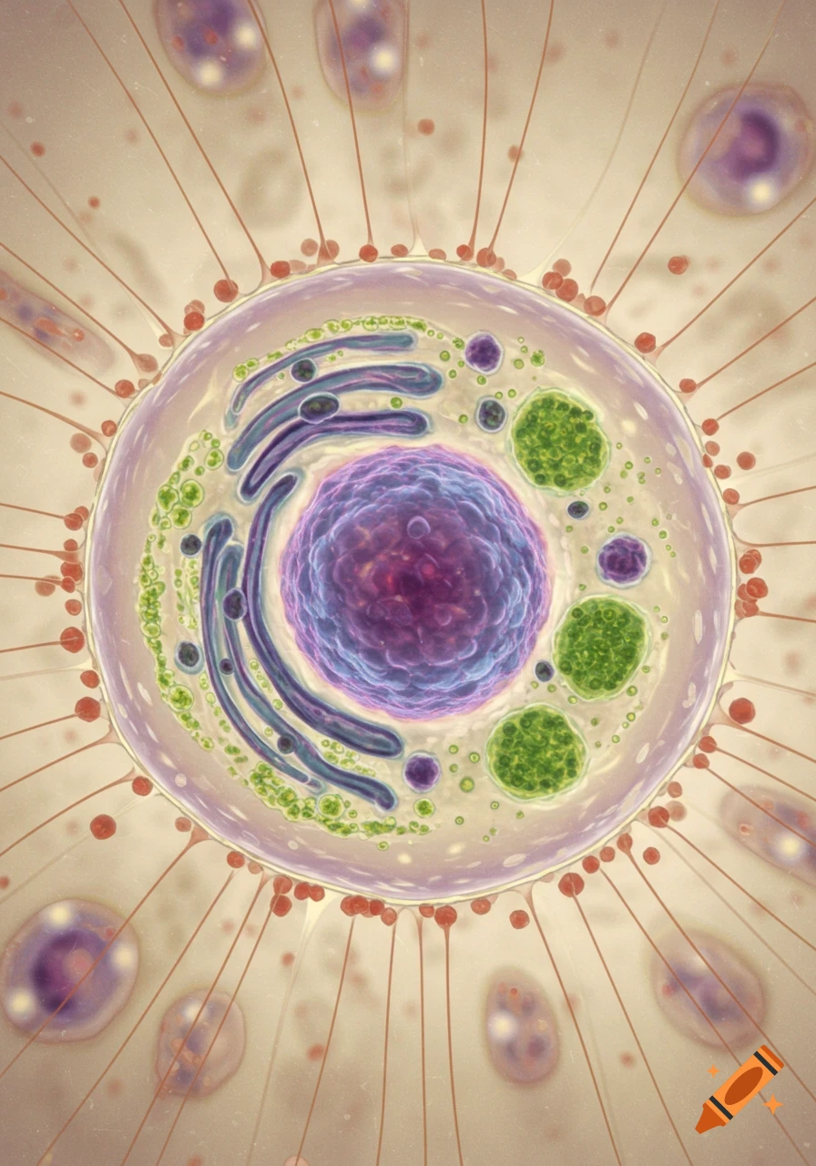
A highly detailed, stylized illustration of a eukaryotic cell with a purple nucleus, blue organelles, and green structures, surrounded by fine red filaments.
Zoom-Aktion
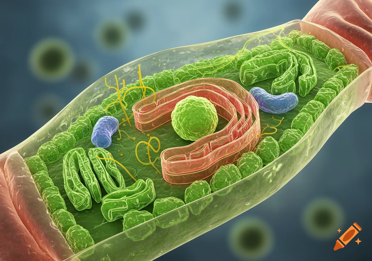
3D rendering of a plant cell with visible organelles like the nucleus, Golgi apparatus, and chloroplasts.
Zoom-Aktion
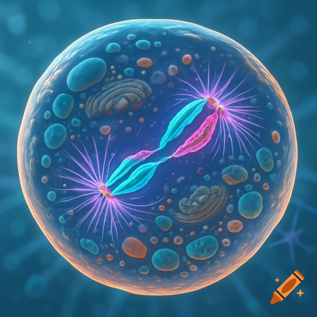
A vibrant microscopic illustration of an animal cell undergoing mitosis, with glowing blue and pink chromosomes separating.
Zoom-Aktion
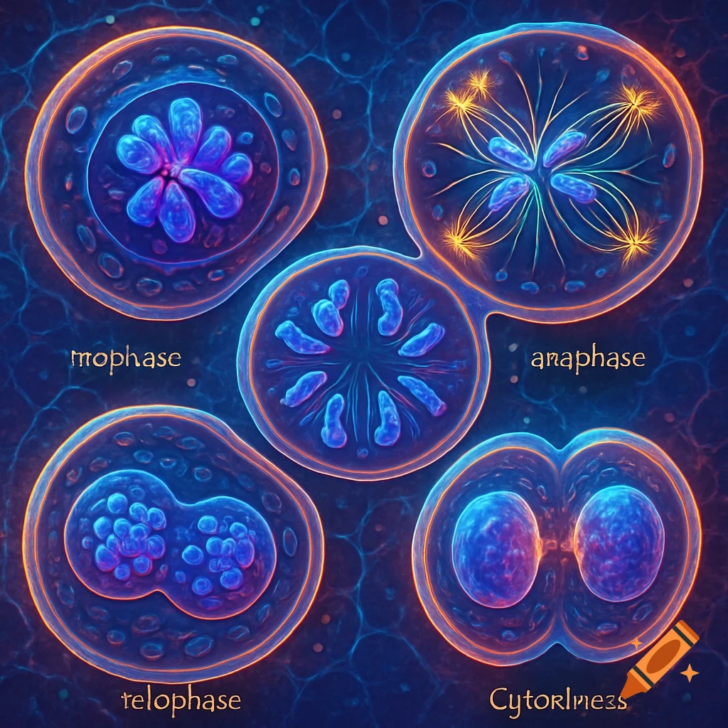
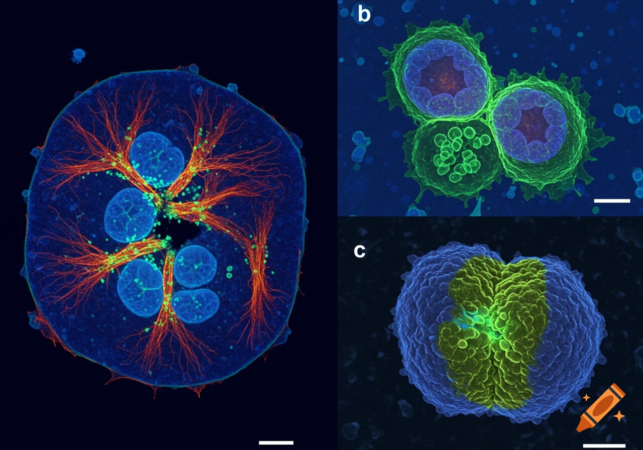
Fluorescent micrographs of animal cells during mitosis and cytokinesis, showing detailed internal structures and processes with various colors.
Zoom-Aktion
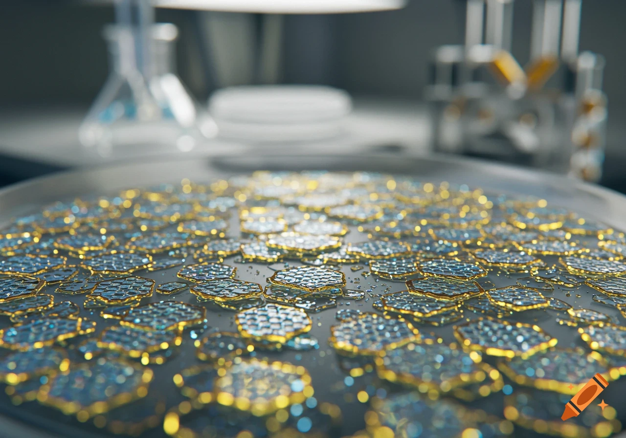
Close-up of shimmering hexagonal nanoplatelets on a surface in a blurred laboratory setting, photorealistic.
Zoom-Aktion
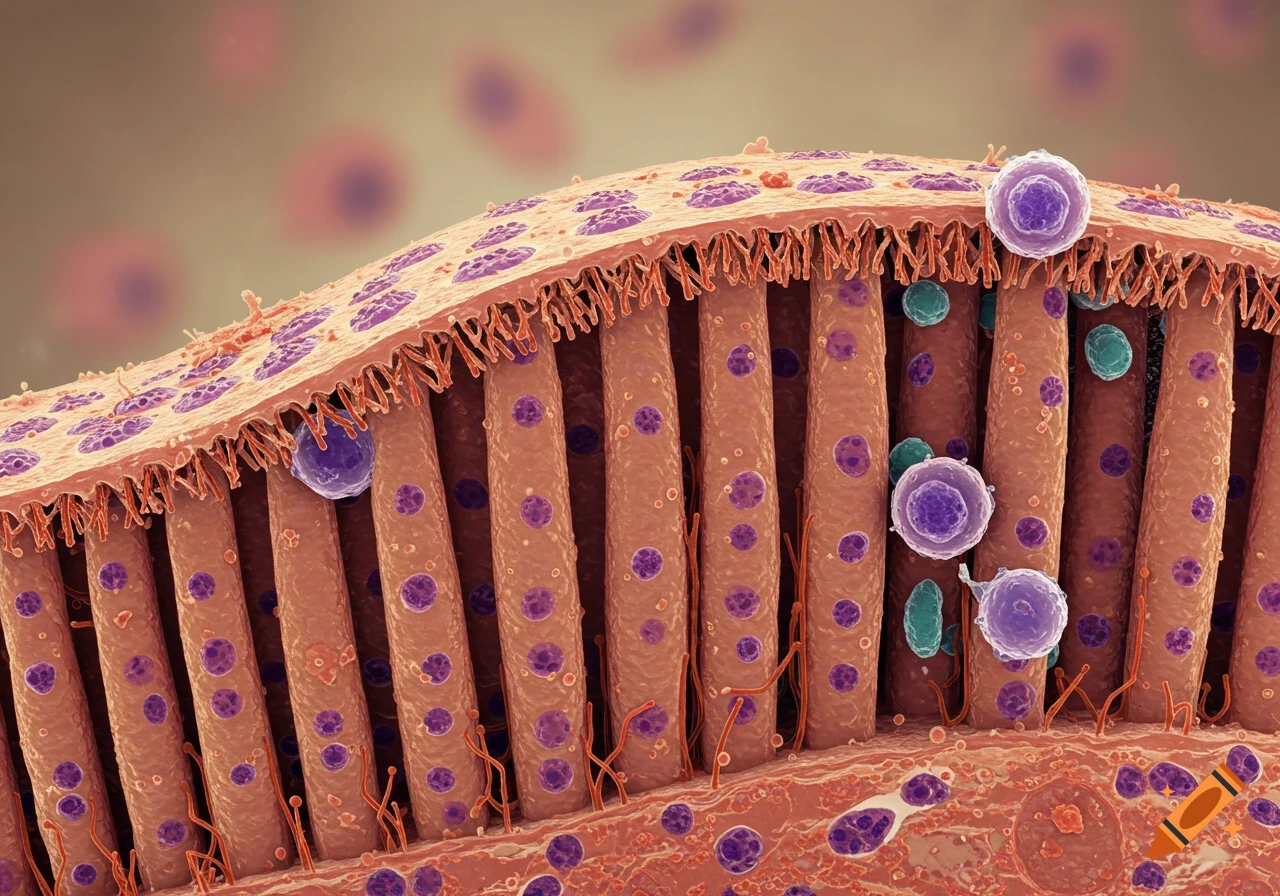
Photorealistic microscopy view of human intestinal villi with columnar epithelial cells and microvilli.
Zoom-Aktion
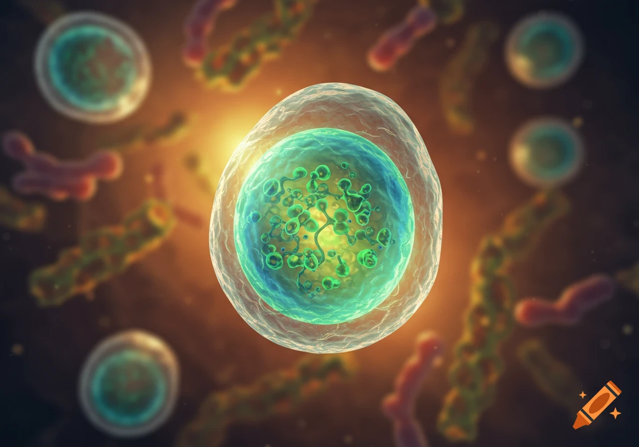
A detailed microscopic view of a translucent cell with green internal structures, surrounded by smaller colorful organisms on a dark, glowing background.
Zoom-Aktion
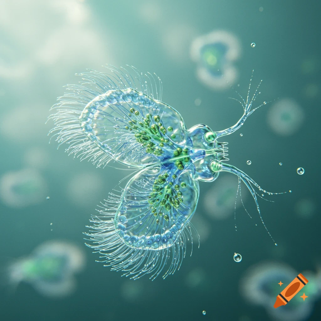
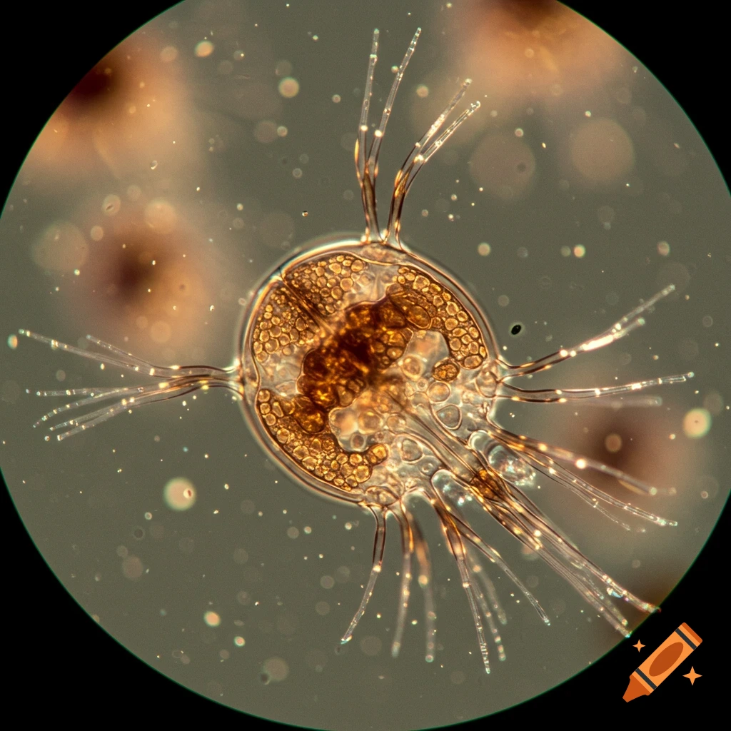
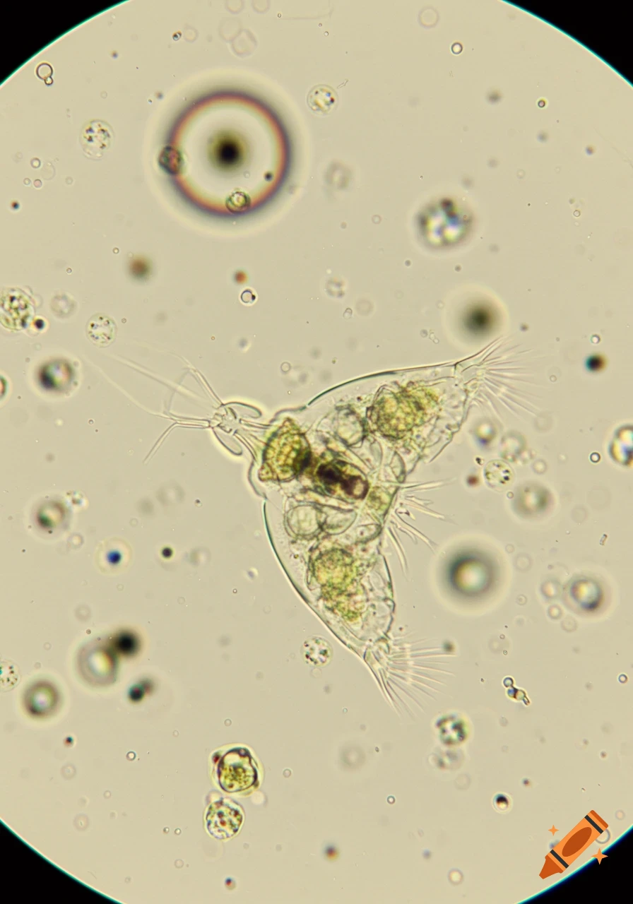
A detailed microscopic view of a translucent rotifer-like organism with many fine cilia and green internal structures, in a watery environment.
Zoom-Aktion
A translucent brown microscopic organism with radiating appendages, seen through a microscope.
Zoom-Aktion
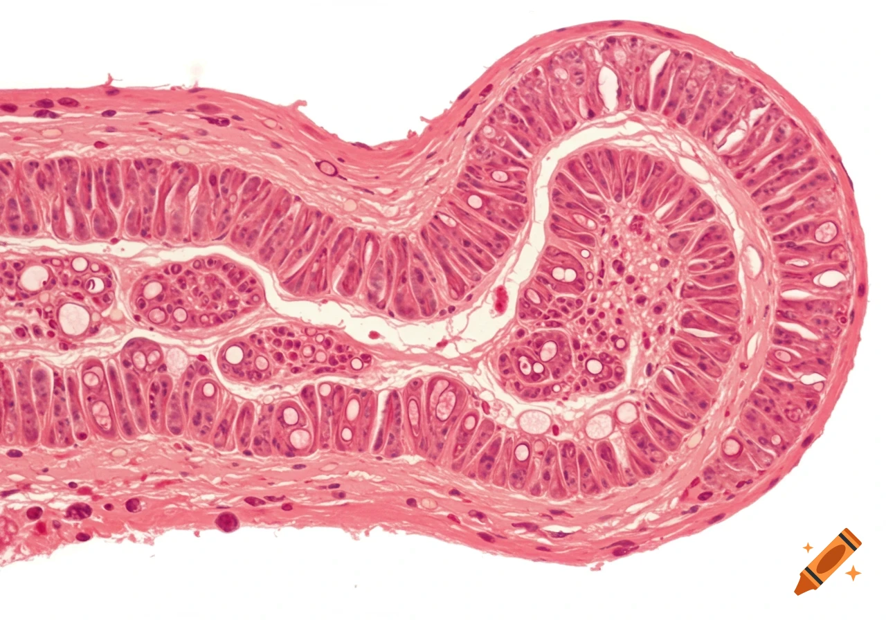
A detailed microscopic image of pink-stained ileum tissue, showing villi, mucosa, and other cellular layers.
Zoom-Aktion
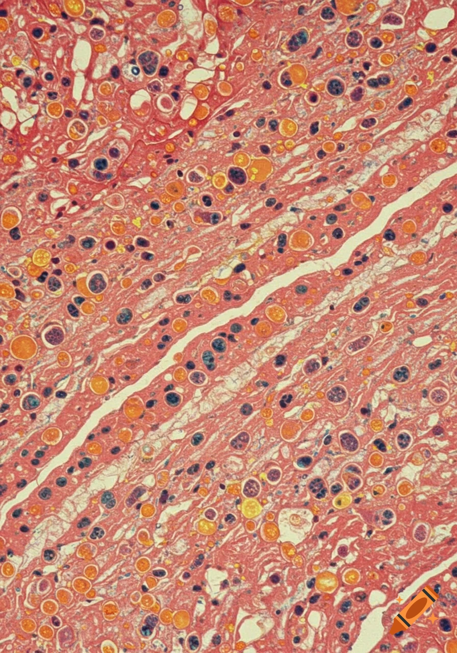
Microscopic view of tissue, possibly cartilage, showing a pink fibrous matrix with scattered round cells in shades of orange, blue, and black.
Zoom-Aktion
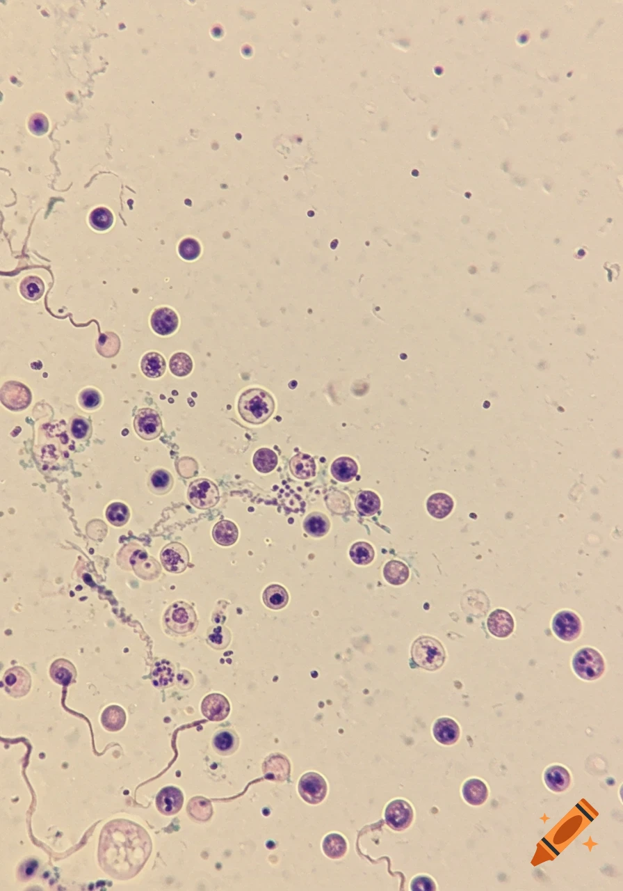
Microscopic view of a canine blood smear showing various types of white and red blood cells.
Zoom-Aktion
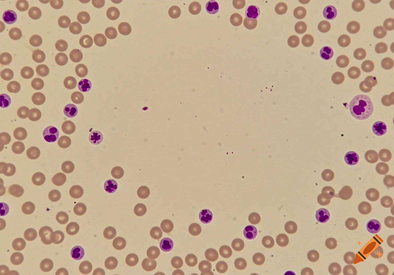
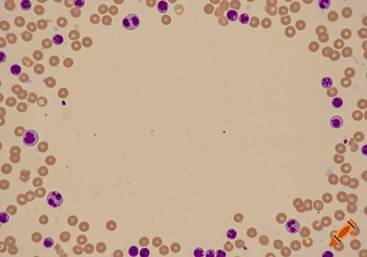
Microscopic view of a canine blood smear showing various blood cells, including red blood cells and different white blood cells, bordering a clear central area.
Zoom-Aktion
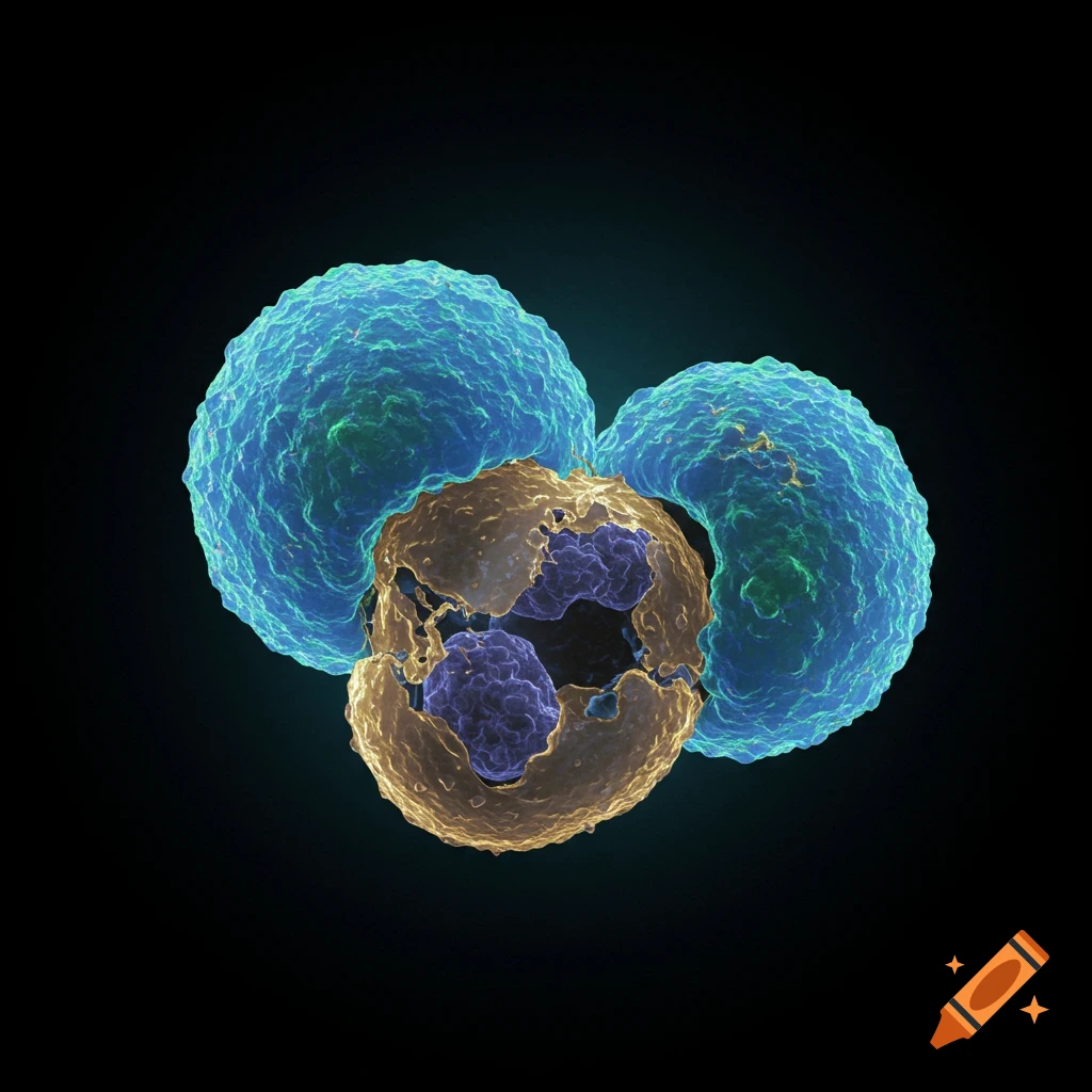
Microscopic view of natural killer cells interacting with a dead cell.
Zoom-Aktion
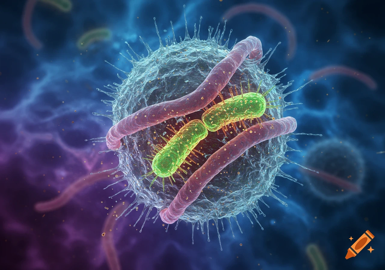
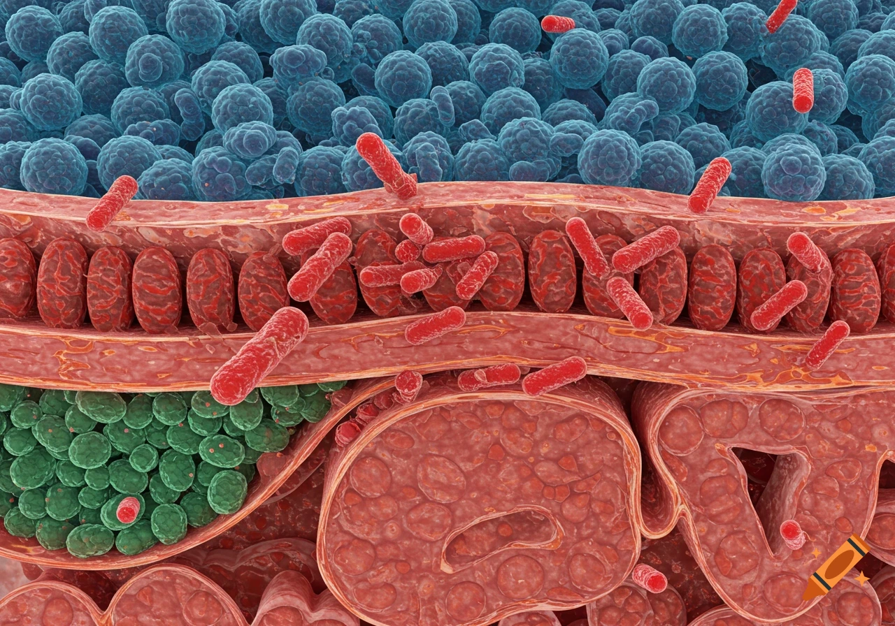
Microscopic view of red rod-shaped bacteria interacting with blue and red cellular structures.
Zoom-Aktion
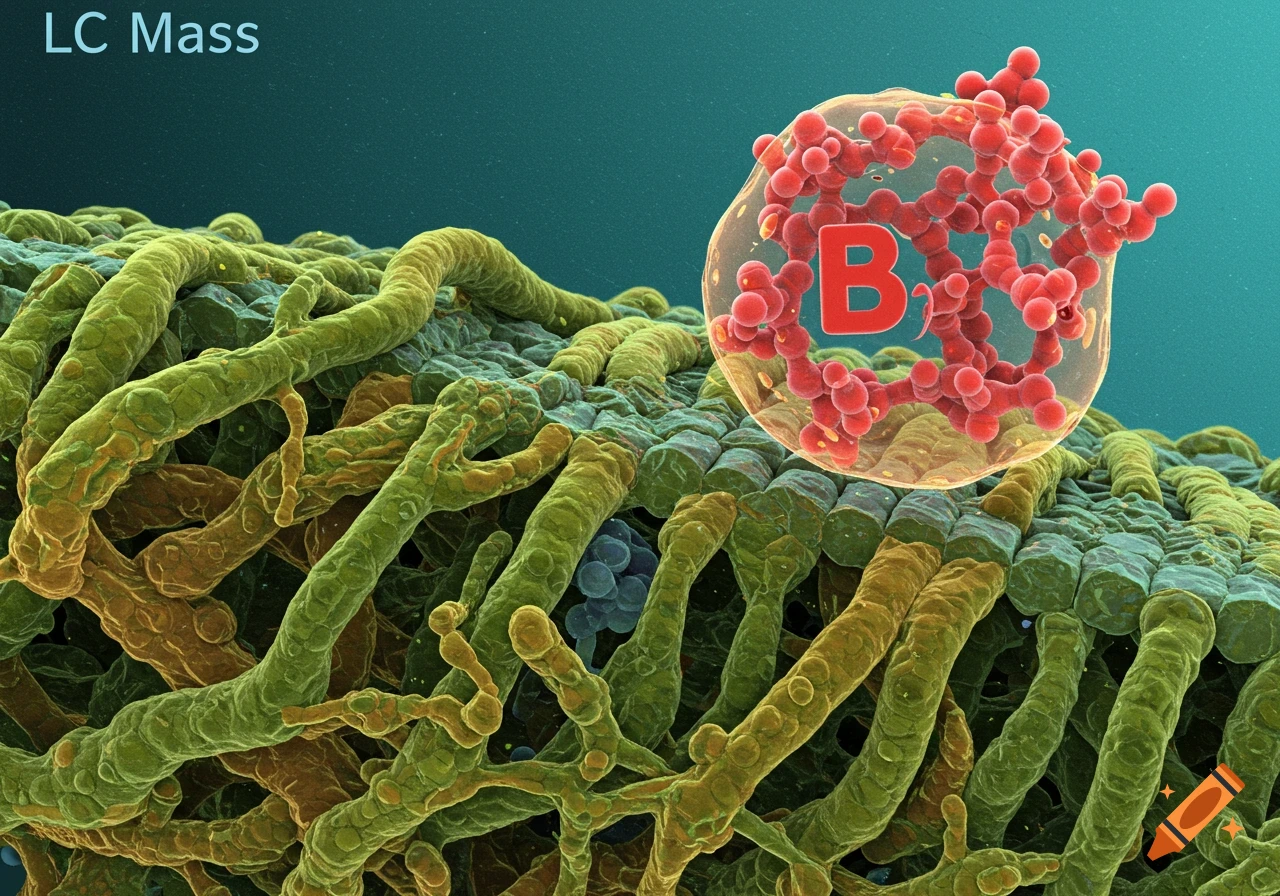
Detailed microscopic view of plant tissue with a cell containing a molecular structure labeled B.
Zoom-Aktion
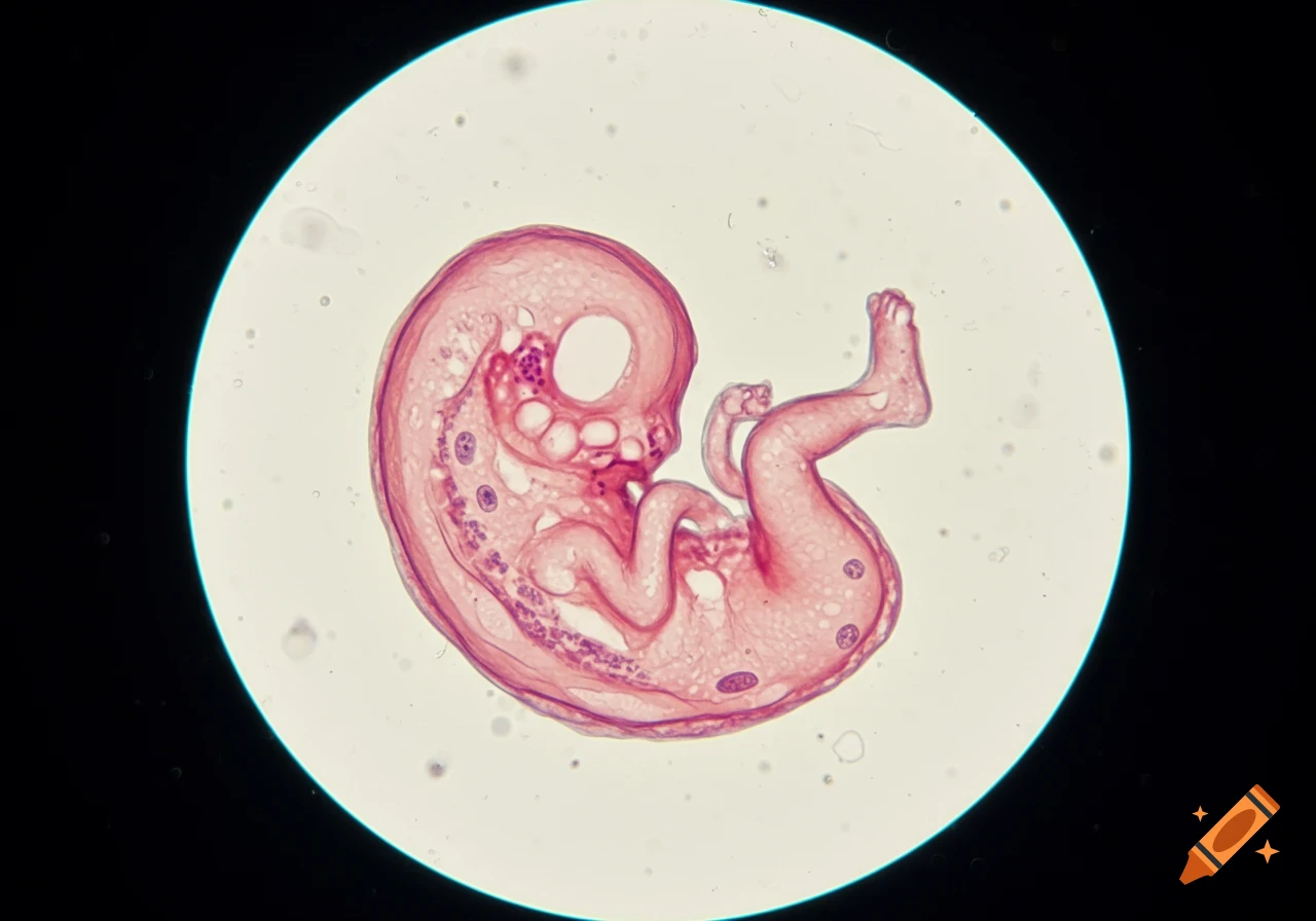
Embryo viewed under a microscope
Zoom-Aktion
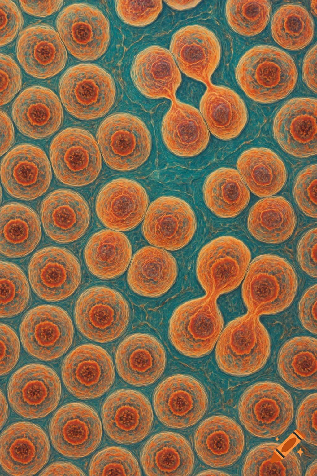
Close-up view of orange biological cells dividing on a blue background.
Zoom-Aktion
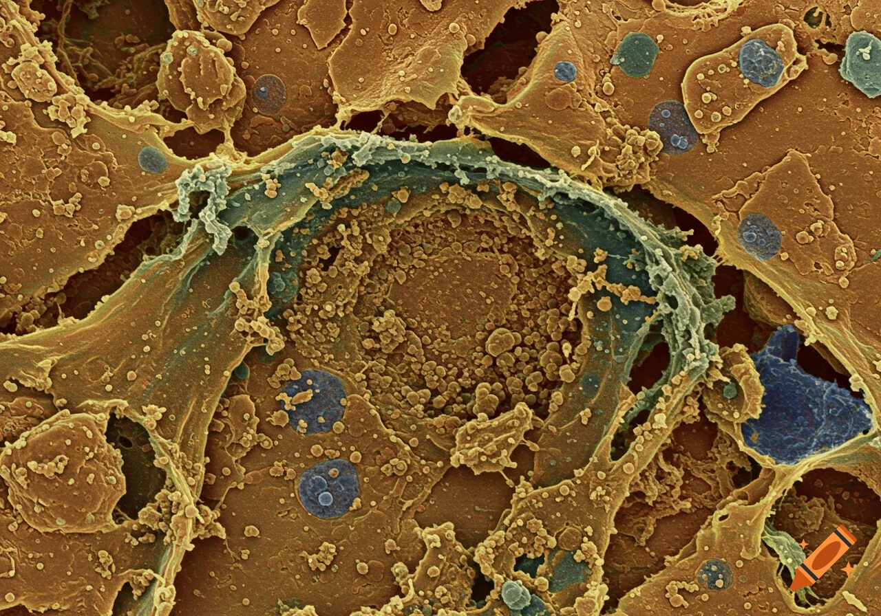
Close-up microscopic view of biological cells and tissue showing detailed textures and forms in brown, orange, blue, and grey.
Zoom-Aktion
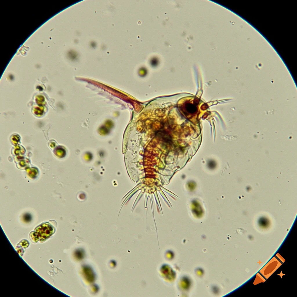
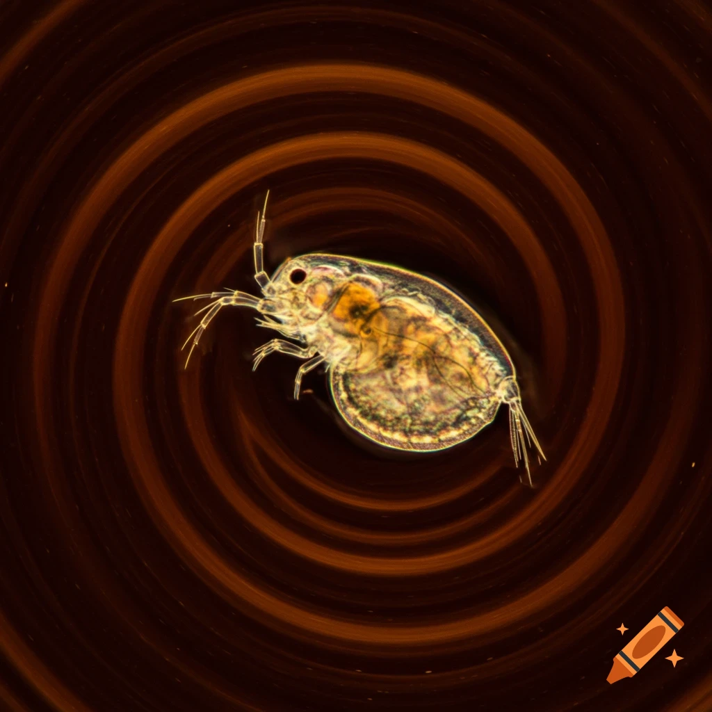
Microscopic daphnia against a swirling brown background
Zoom-Aktion
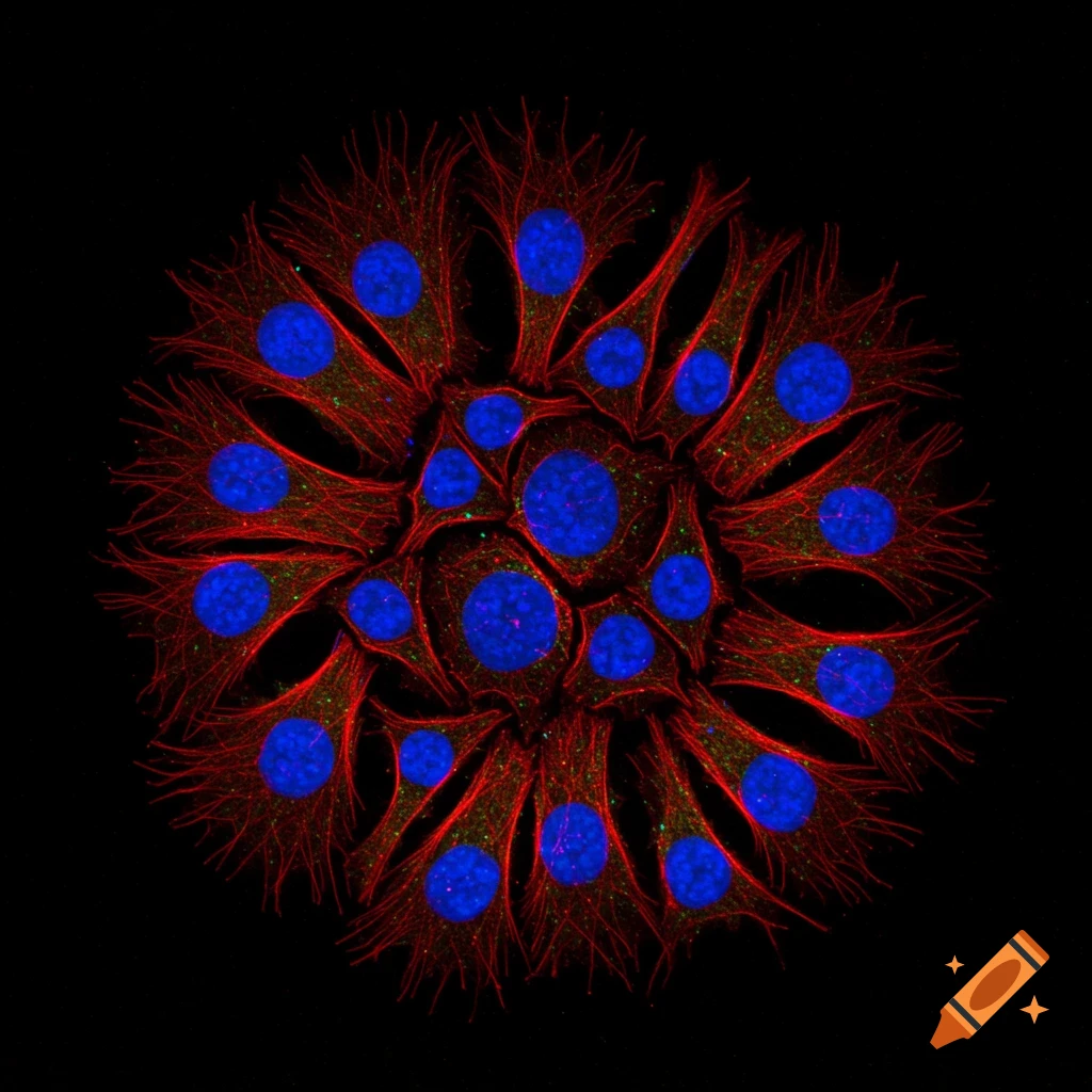
Microscopic view of cells with blue nuclei and red cytoskeleton on a black background.
Zoom-Aktion
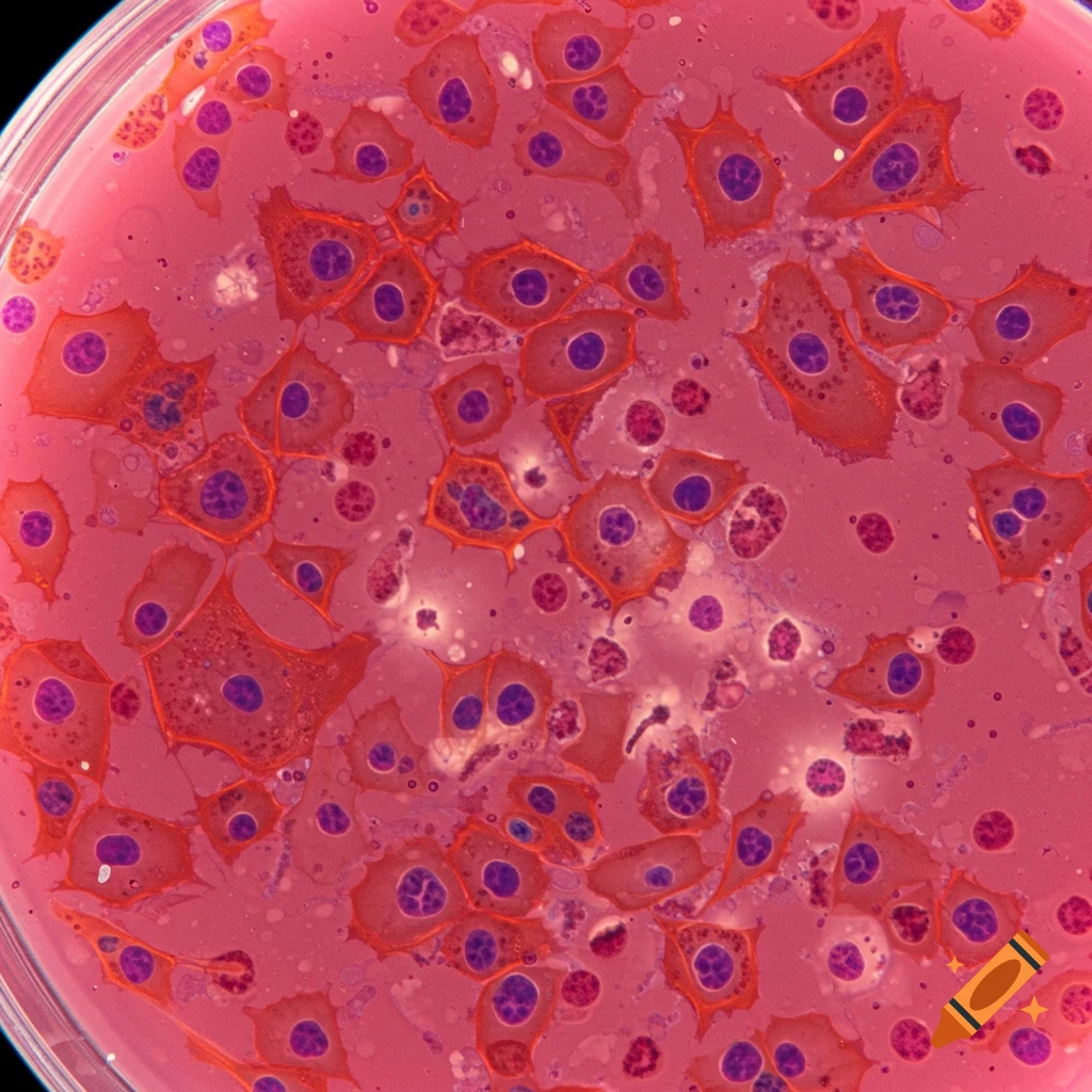
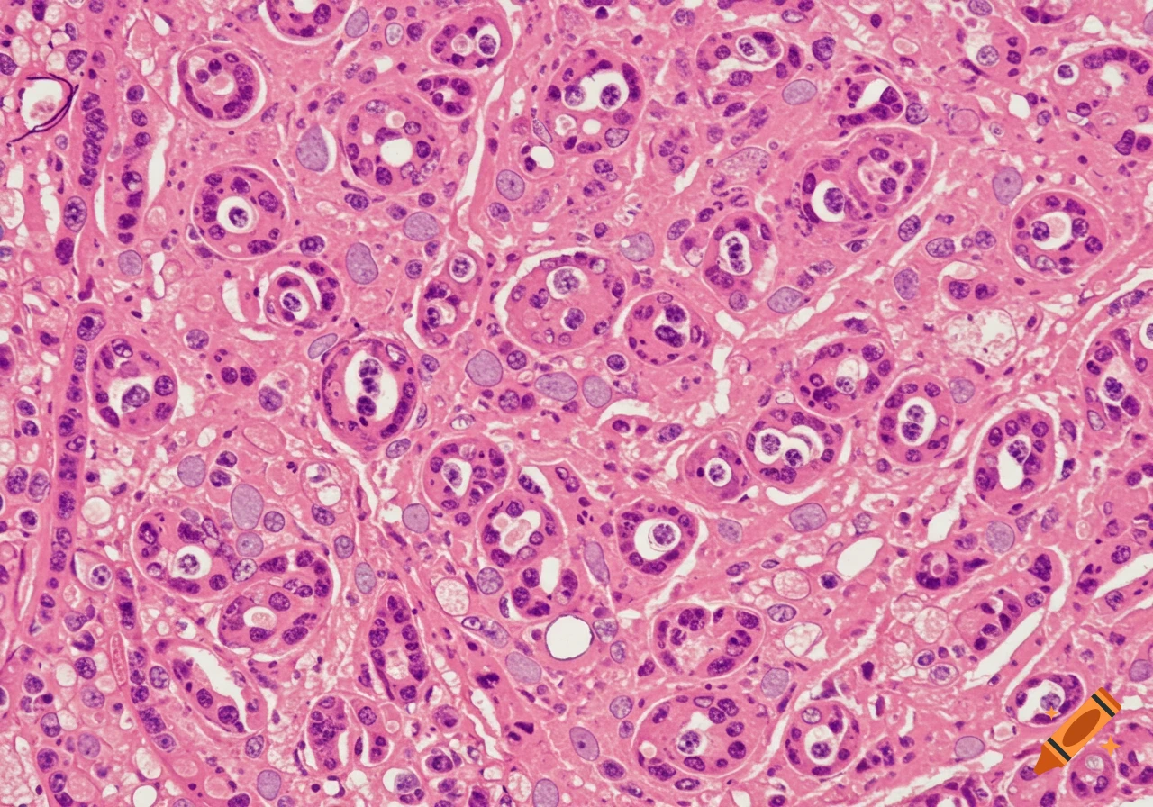
Microscopic view of mammalian cells in a petri dish, stained pink with purple nuclei
Zoom-Aktion
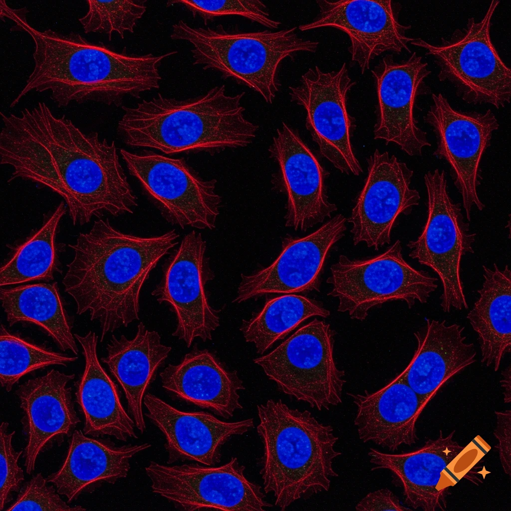
A microscopic view of numerous fluorescently stained cells with blue nuclei and red cytoplasm on a black background.
Zoom-Aktion
Immunofluorescence images of GAP-43 staining showing axonal regrowth post-tumor resection
Zoom-Aktion
Microscopic view of ketchup, mustard, and dirt mixture
Zoom-Aktion
Microscopic view of dry amoeba with interconnected organs
Zoom-Aktion
Parvovirus B19 microscopic image
Zoom-Aktion
Microscopic view of chromatin in a cell's nucleus
Zoom-Aktion
Long range microscopy on a drone
Zoom-Aktion
Genetics and electron microscope in high definition
Zoom-Aktion
Genetics and electron microscope in high definition
Zoom-Aktion
Earth zoom under microscope
Zoom-Aktion
Microscope image of red and white blood cells
Zoom-Aktion
Microscopic view of a cell with red-black gradient dot
Zoom-Aktion
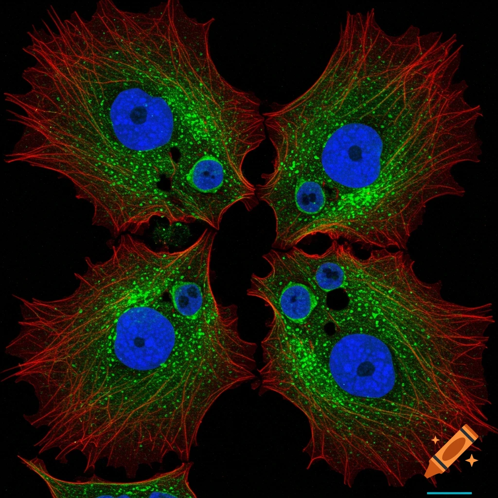
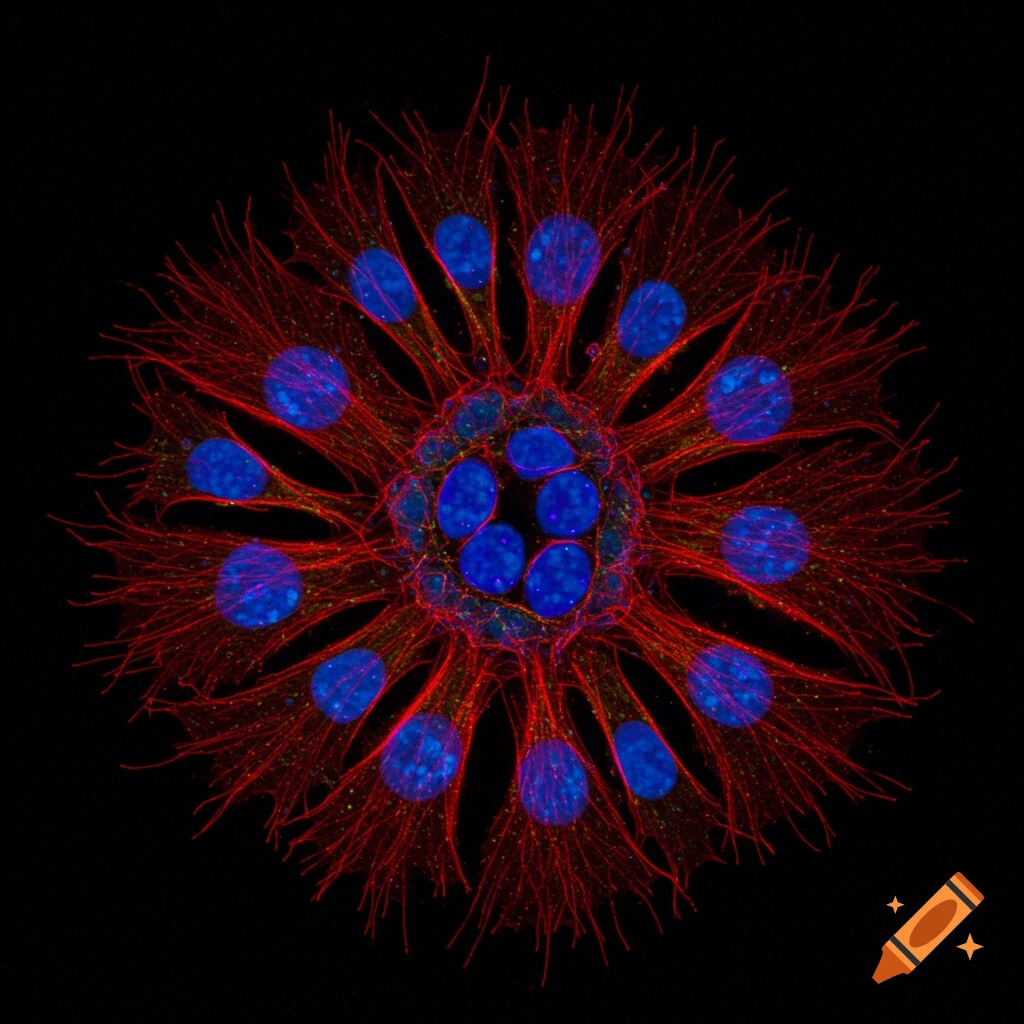
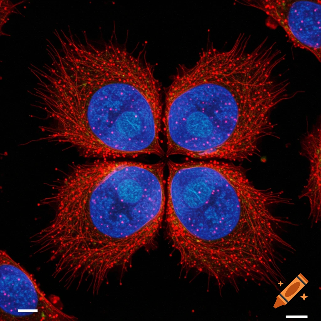
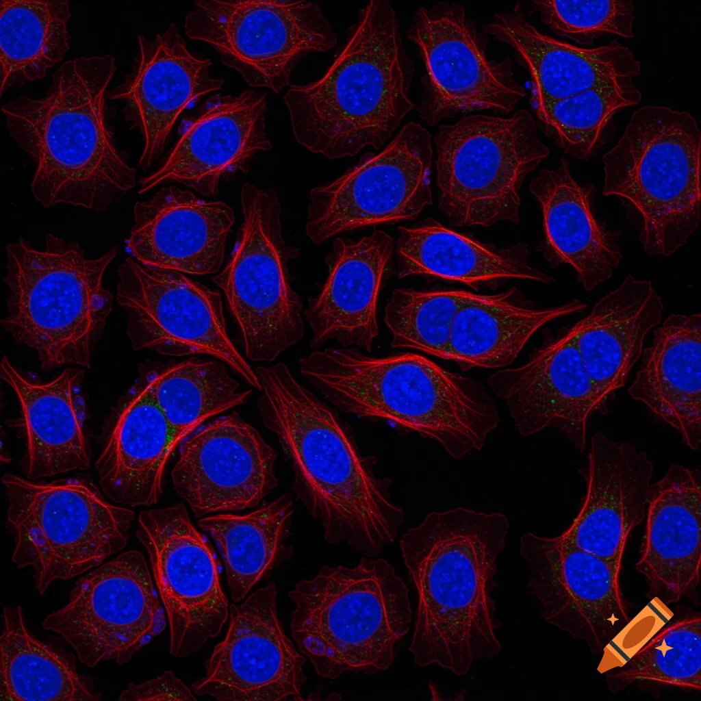
COS7 cells showing actin in green, golgi in red, and nucleus in blue under fluorescent microscopy
Zoom-Aktion
Fluorescent gene transcript in cells of tissue in 3D
Zoom-Aktion
Magnified intricate fungal structures in a culture petri dish
Zoom-Aktion
Copper structure analyzed by nanoscale microscopy
Zoom-Aktion
Extreme close up of bright yellow-green citrus fruit skin under a microscope
Zoom-Aktion
High magnification of a translucent membrane in black and white
Zoom-Aktion
High magnification of translucent membrane with histological stains
Zoom-Aktion
Microscopic view of algae
Zoom-Aktion
Microscopic view
Zoom-Aktion
Microscopic view
Zoom-Aktion
Cell
Zoom-Aktion
Genetics and electron microscopy exploring the intricate tapestry of life at a hyperdetailed level with 3D renders
Zoom-Aktion
Genetics and electron microscopy exploring the intricate tapestry of life at a hyperdetailed level with 3D renders
Zoom-Aktion
Detailed drawing of an amoeba on white background in HD
Zoom-Aktion
Microscopic view of a brain
Zoom-Aktion
monocyte on white background
Zoom-Aktion
seamless pattern of human lung cells under a microscope
Zoom-Aktion
microscopic view of hair strands
Zoom-Aktion
logo combining electron microscopy and a coffee bean
Zoom-Aktion
close-up of metal mesh separating gas flow
Zoom-Aktion
microscopic image of cells undergoing mitosis
Zoom-Aktion
microscopy image of carbon nanotubes on carbon fibre
Zoom-Aktion
fractal fungal growth inspired by Haeckel
Zoom-Aktion
fractal fungal growth inspired by Haeckel
Zoom-Aktion
close-up of connective tissue under a microscope
Zoom-Aktion
cross-section of plant stem under a microscope
Zoom-Aktion
8-bit style microscopy science photography
Zoom-Aktion
microscopic view of a crystal diamond texture
Zoom-Aktion
microscopic view of a crystal diamond texture
Zoom-Aktion
microscopic image of virus being engulfed
Zoom-Aktion
close-up of a peridot crystal under a microscope
Zoom-Aktion
scientist observing a violet flower under a microscope
Zoom-Aktion
black and white microscope image of a rhododendron stigma flower
Zoom-Aktion
black and white microscopy of a buddleja flower
Zoom-Aktion
black and white scanning electron microscopy image of a plastic snowflake
Zoom-Aktion
scanning electron microscopy of lichen cells
Zoom-Aktion
black and white SEM image of borage
Zoom-Aktion
black and white microscopy image of lichen
Zoom-Aktion
black and white microscopic image of tree bark
Zoom-Aktion
black and white image of flower petals under electron microscope
Zoom-Aktion
scanning electron microscopy image of black and white pollen
Zoom-Aktion
fluorescence microscopy image
Zoom-Aktion
view of human gallbladder cells under the microscope
Zoom-Aktion
microscopy of human uterine cells
Zoom-Aktion
microscopic view of human thyroid cells
Zoom-Aktion
microscopic view of human fat cell
Zoom-Aktion
microscopic view of human oral mucosa cells
Zoom-Aktion
microscopic view of human kidney cells
Zoom-Aktion
microscopic view of human intestinal cells
Zoom-Aktion
microscopic view of a human bone cell
Zoom-Aktion
microscopic view of human muscle cells
Zoom-Aktion
high-resolution image of microscopic particles shaped as crosses
Zoom-Aktion
microscopic image of a bumpy animal cell
Zoom-Aktion
microscopic image of a bumpy animal cell
Zoom-Aktion
Scientific illustration of bullet-shaped purple rabies virions with spikes on a glowing teal neuron axon.
Zoom-Aktion
Close-up, detailed view of a single cell or spore with a clear outer membrane, floating in a liquid with other blurred cells in the background.
Zoom-Aktion
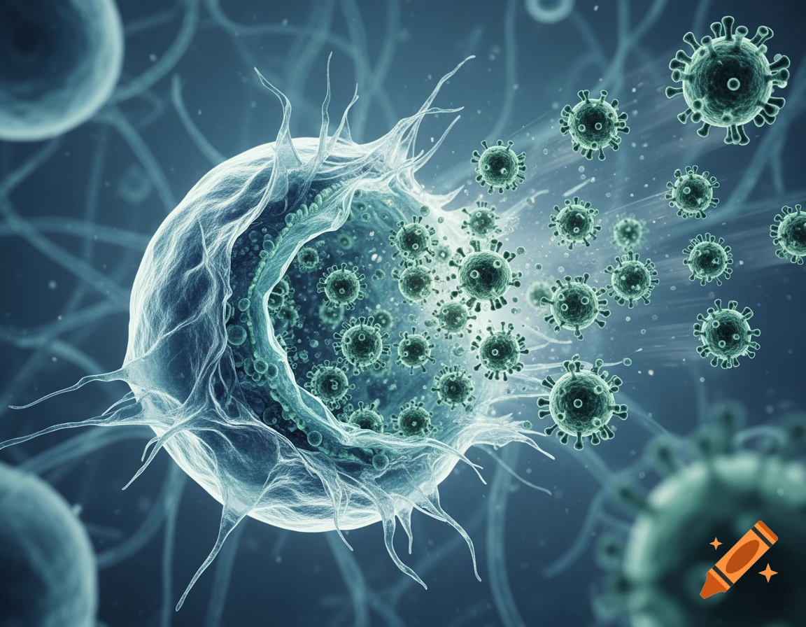
A large cell bursting and releasing numerous spikey virus particles in a dark teal microscopic setting.
Zoom-Aktion
A vibrant biological cell cross-section, glowing blue, purple, and green organelles on a dark blue background in fluorescent microscopy style.
Zoom-Aktion
Microscopic view of glowing blue and silver microspheres surrounded by greenish cell-like structures.
Zoom-Aktion
Detailed microscopic view of a human fallopian tube cross-section, revealing internal folds and cilia-like structures.
Zoom-Aktion
Photorealistic microscopic cross-section of pink intestinal villi against a blurred laboratory background with a microscope and screen.
Zoom-Aktion
Scanning electron micrograph of a spiky, irregular cell or microorganism with internal granular structures, set against a porous background.
Zoom-Aktion
A detailed 3D rendering of a spherical purple cell covered in green, bulbous protein structures on a blurred dark background.
Zoom-Aktion
Microscopic view of a translucent, elongated trypanosome-like organism filled with blue and red spherical particles against a blurred reddish background.
Zoom-Aktion
Photorealistic close-up of a scanning electron microscope's sample chamber with a copper component, SE, BSE, and EDX detectors.
Zoom-Aktion
A microscopic view of a vibrant blue and green cell with a nucleus and organelles.
Zoom-Aktion
Macroscopic view of a translucent, yellowish-green organic specimen with intricate patterns, seen through a stereomicroscope.
Zoom-Aktion
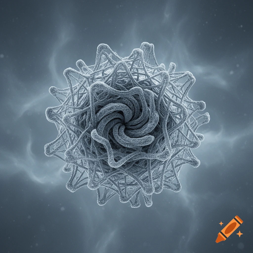
Microscopic grey-blue virus with a swirling core and an interlocking lattice against a textured background.
Zoom-Aktion
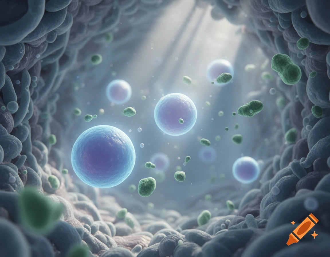
Microscopic view of blue and green cells, likely lymphocytes, floating within a biological environment with light rays.
Zoom-Aktion
Microscopic view of a glowing synapse between two neurons, showing synaptic buttons and a neurotransmitter.
Zoom-Aktion
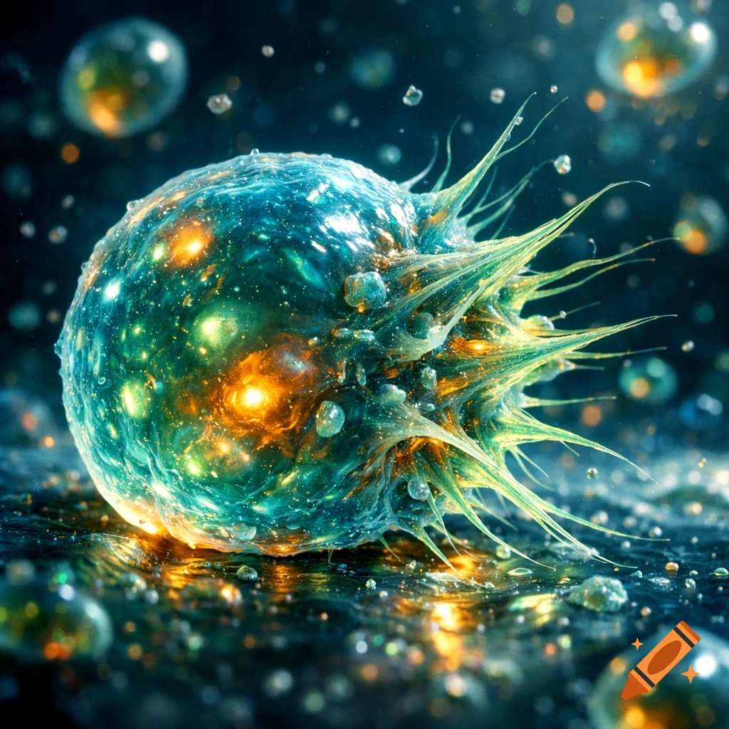
A glowing, spiky, microscopic cell-like organism in vibrant blue and green with orange internal lights, against a dark, blurry background.
Zoom-Aktion
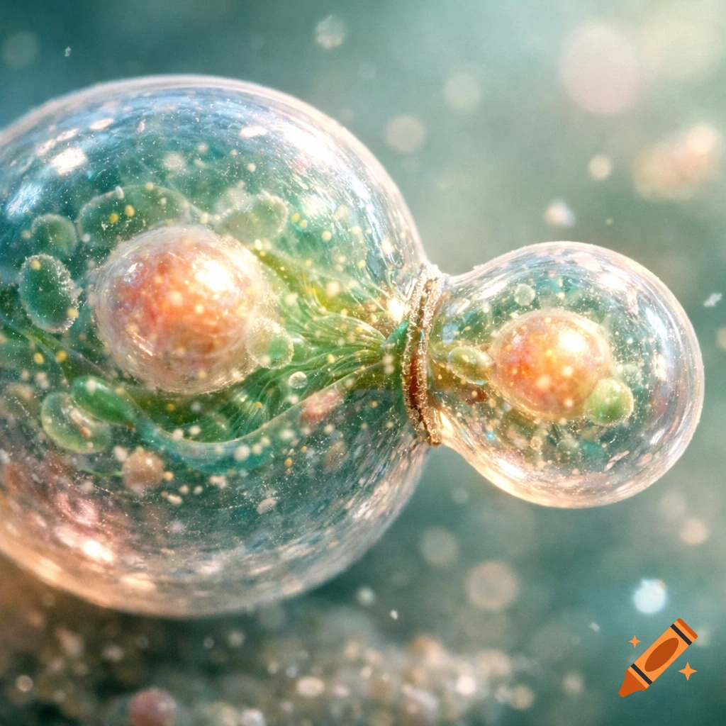
A microscopic view of a large clear cell budding with internal green and orange structures, attached to a smaller transparent cell.
Zoom-Aktion
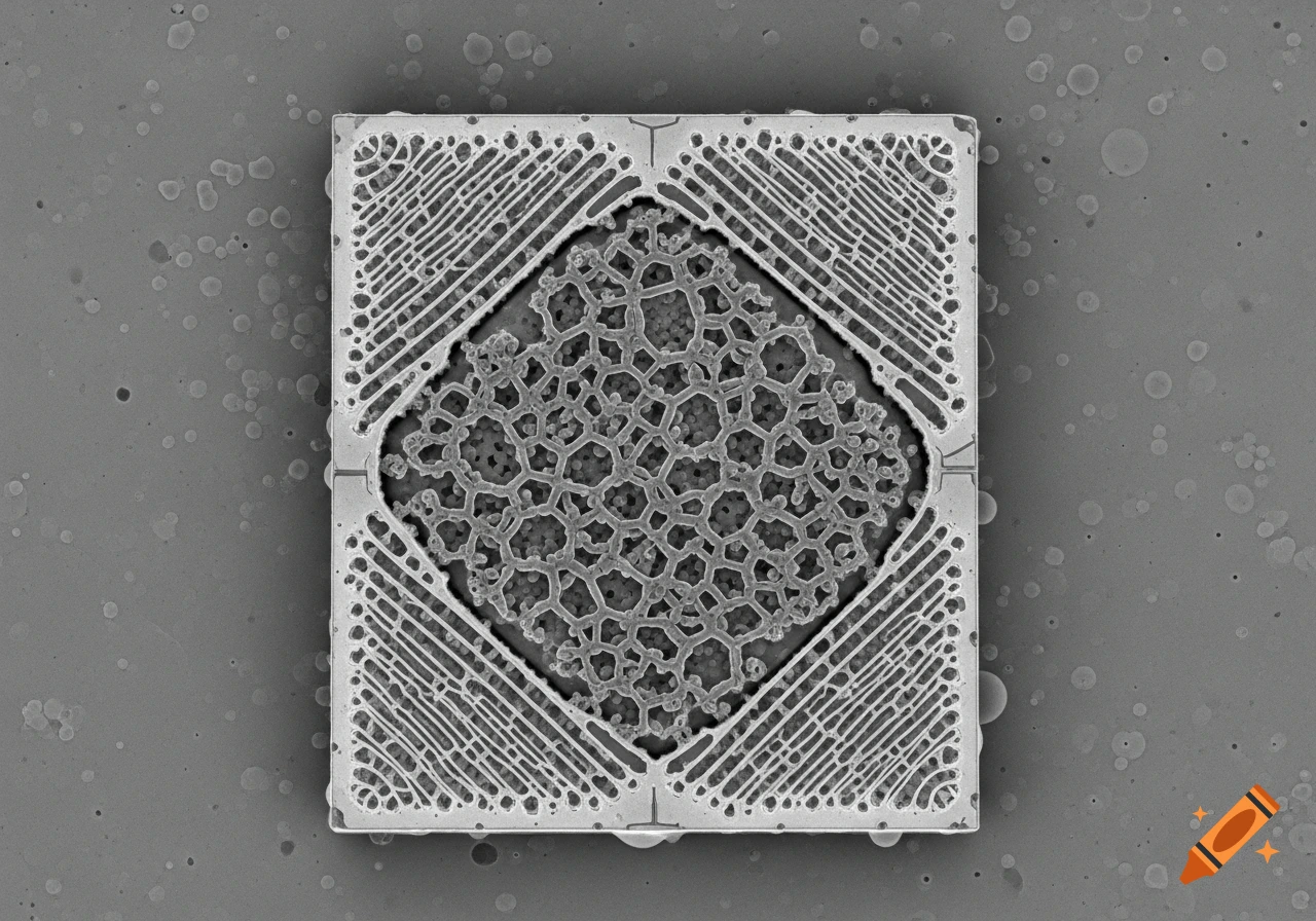
Detailed gray-scale electron micrograph showing intricate square and hexagonal patterns on a microchip.
Zoom-Aktion
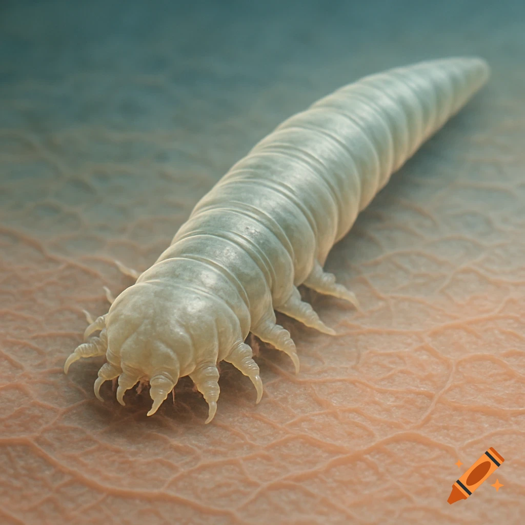
Close-up, detailed view of a light-colored Demodex face mite crawling on textured human skin.
Zoom-Aktion
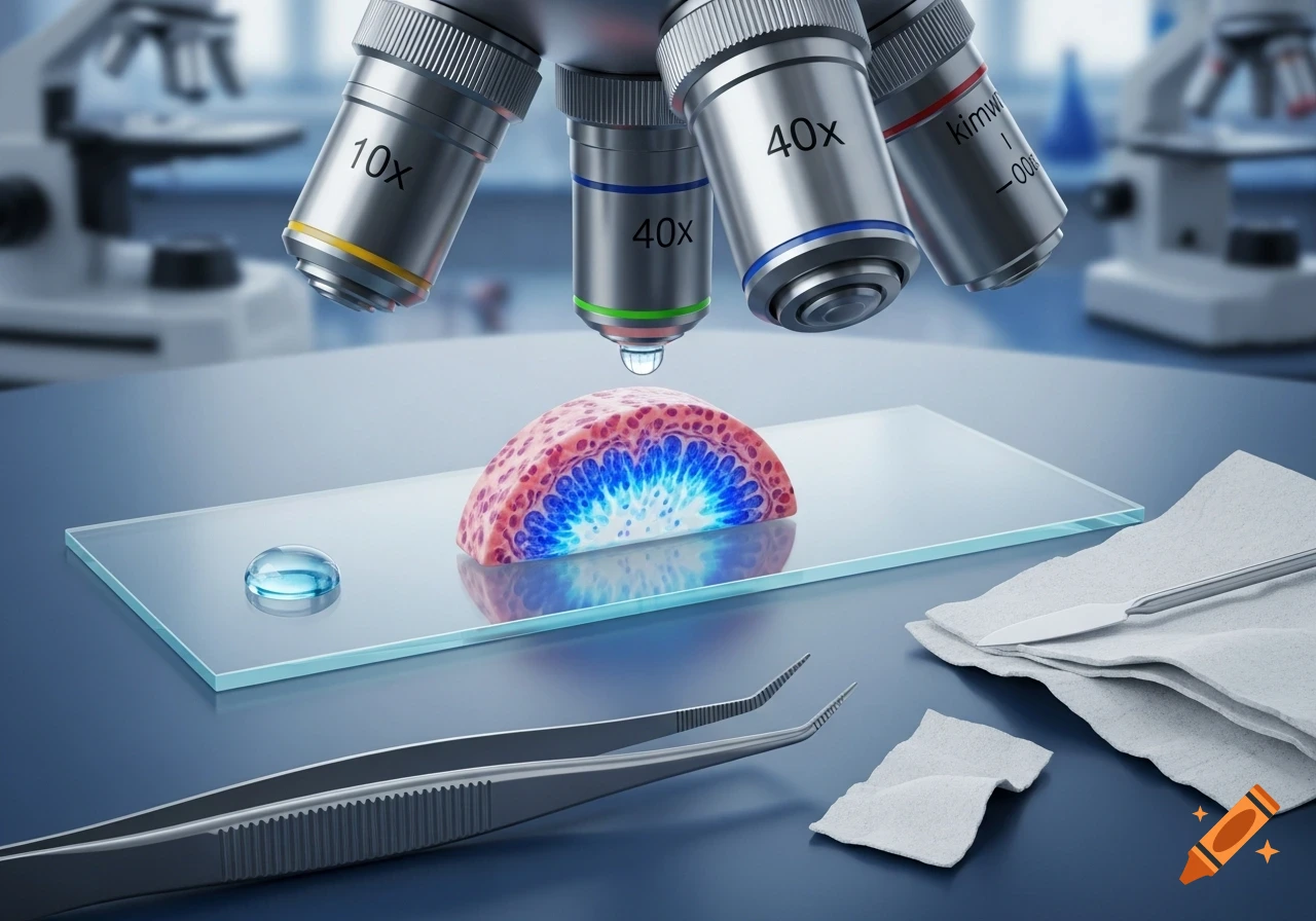
A close-up view of a microscope observing a glowing biological tissue slice on a glass slide. Microscope objectives are visible above.
Zoom-Aktion
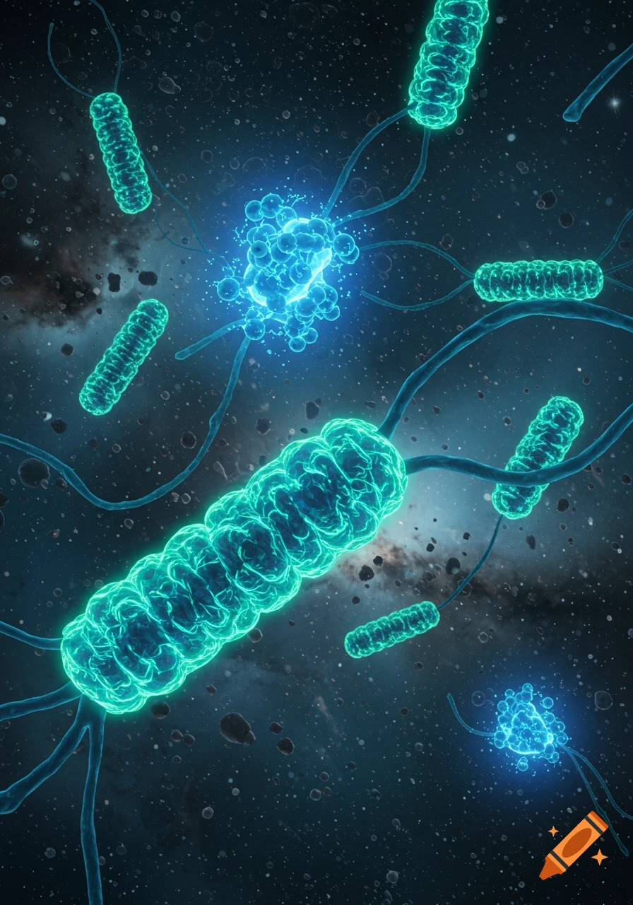
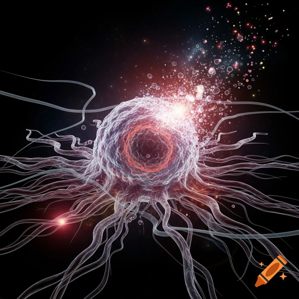
Glowing, tentacled cell dissolving into bright particles against a dark, starry background.
Zoom-Aktion
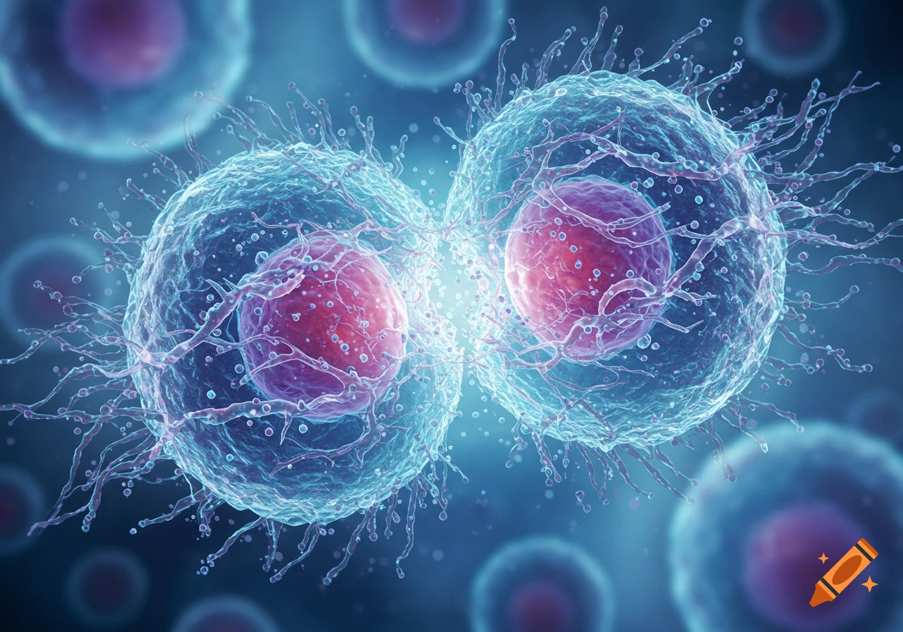
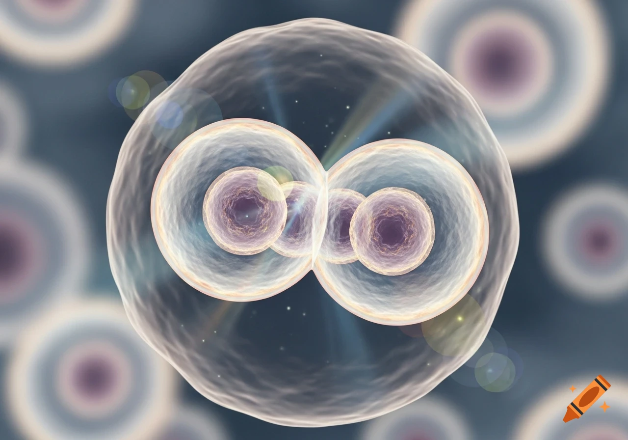
Two translucent, glowing cells with internal nuclei appear to be dividing or fusing, against a blurry cellular background.
Zoom-Aktion
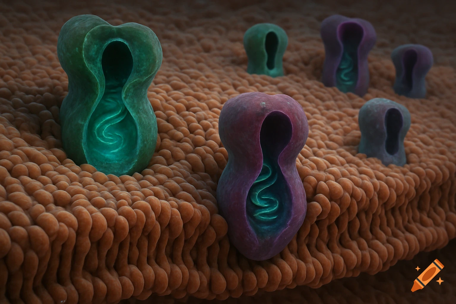
Green and purple protein-like structures embedded in a textured brown biological membrane surface.
Zoom-Aktion
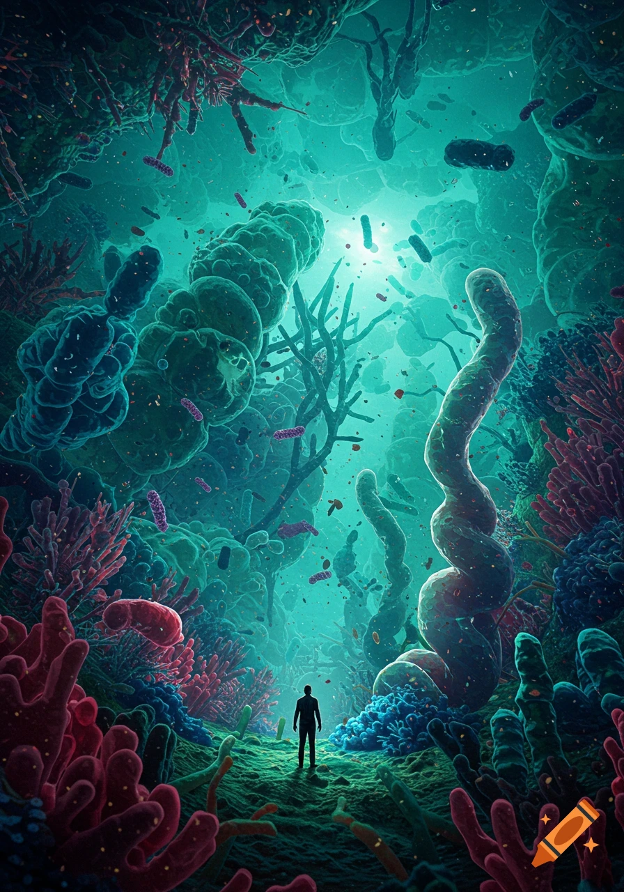
A human silhouette stands amidst a vibrant, otherworldly microscopic landscape of colorful bacteria, cells, and coral-like structures, with light shining from above.
Zoom-Aktion
A cluster of vibrant green, blue, and purple microscopic cells or viruses, some with red nuclei, on a white background.
Zoom-Aktion
A grayscale scanning electron microscope (SEM) image of a cube-shaped salt crystal.
Zoom-Aktion
Highly detailed microscopic view of a translucent, spiky cell or virus with dark specks against a dark background.
Zoom-Aktion
A microscopic view of a cell with a central violet nucleus and transparent cytoplasm.
Zoom-Aktion
A close-up, highly detailed microscopic view of a vibrant purple cell with internal structures, against a dark background.
Zoom-Aktion
A glowing blue-green microscopic cell with intricate internal structures like DNA and organelles against a dark background.
Zoom-Aktion
A fluorescence microscopy image of a lymph node section showing red B cells, green T cells, and blue cell nuclei.
Zoom-Aktion
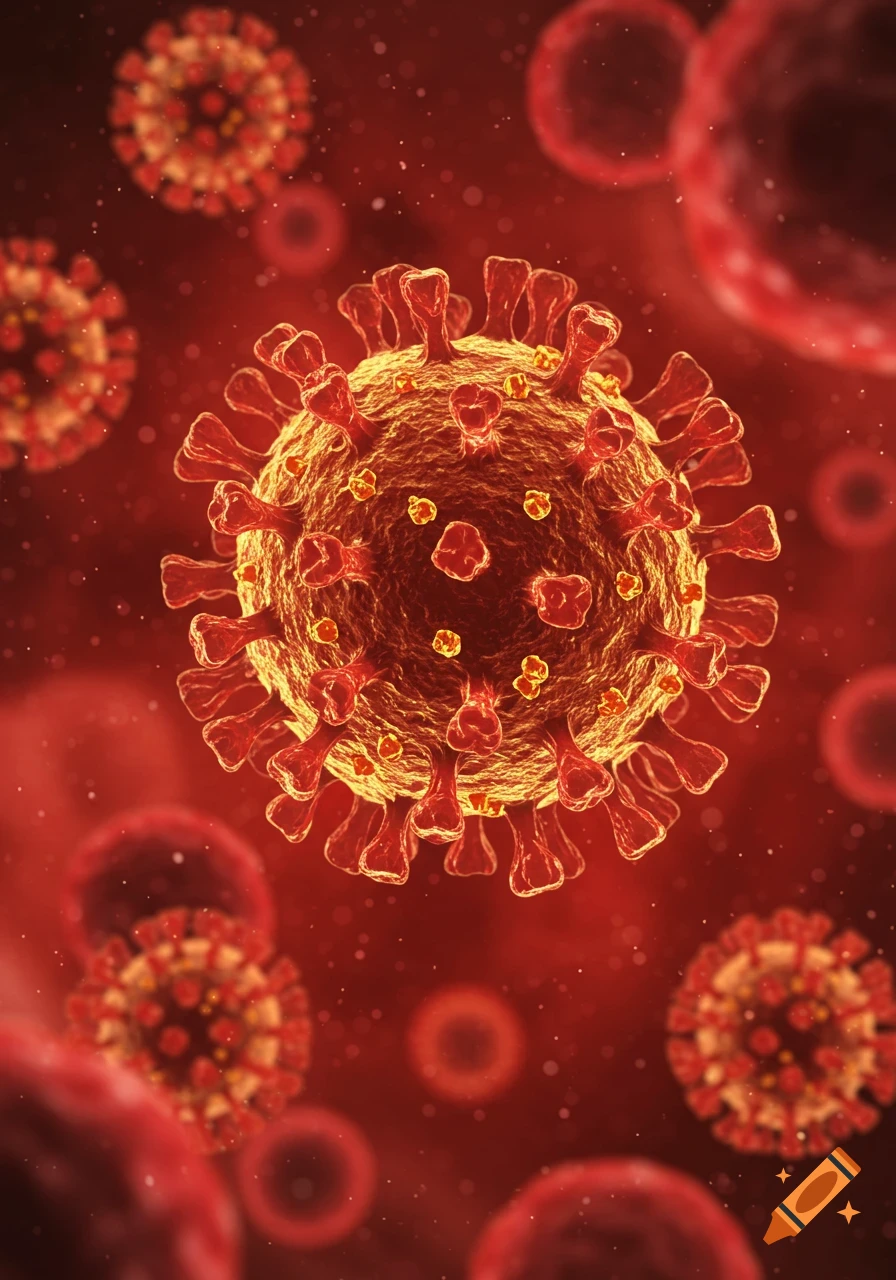
Detailed 3D illustration of a SARS-CoV-2 virus, reddish-orange with spike proteins, against a dark red background.
Zoom-Aktion
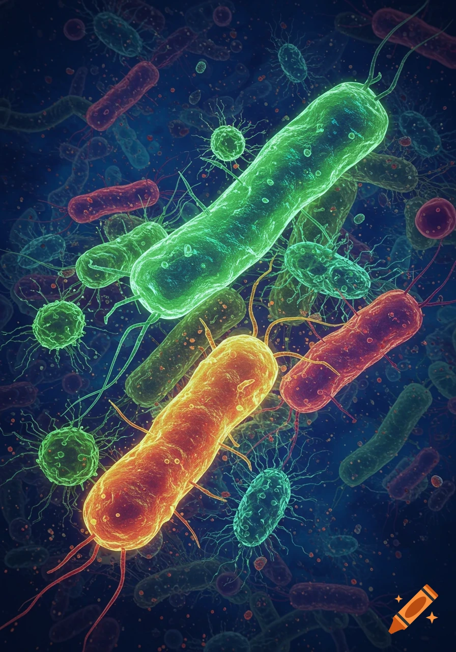
A vibrant, microscopic view of multiple glowing green, orange, and red bacteria and various microorganisms floating in a dark blue fluid.
Zoom-Aktion
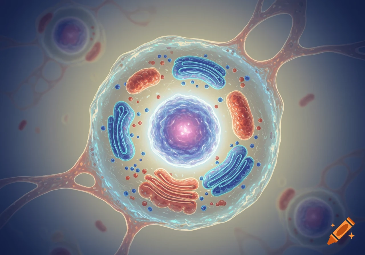
A detailed 3D rendering of a biological cell with a glowing nucleus, mitochondria, and other organelles.
Zoom-Aktion
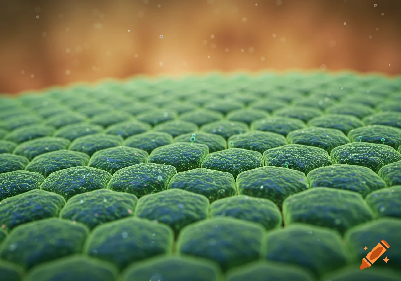
Microscopic close-up of green epithelial cells with a textured surface, against a blurred brown background.
Zoom-Aktion
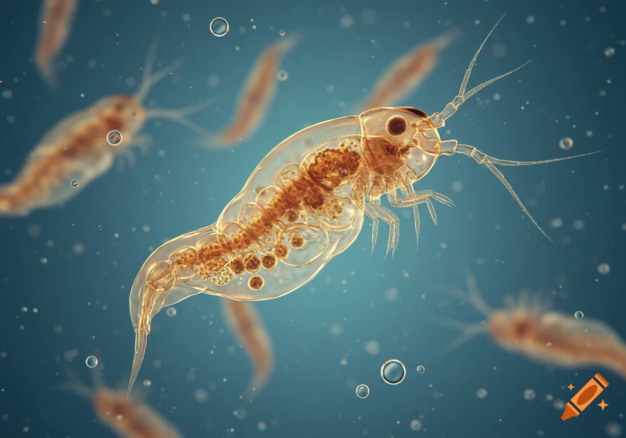
Close-up microscopic view of a translucent brown rotifer with visible internal organs and bubbles, against a blue background.
Zoom-Aktion
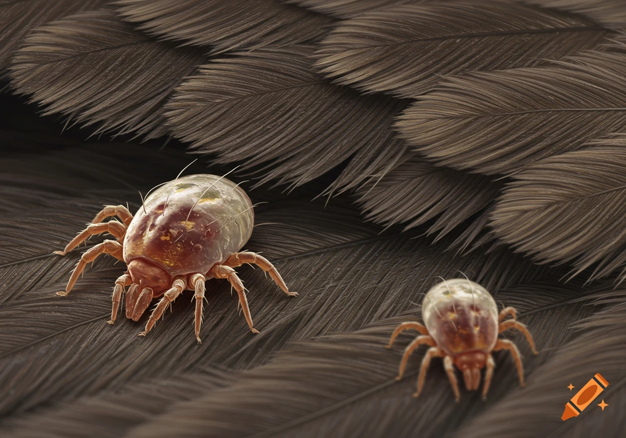
Two detailed microscopic bird mites are shown on a textured brown surface, appearing like feathers.
Zoom-Aktion
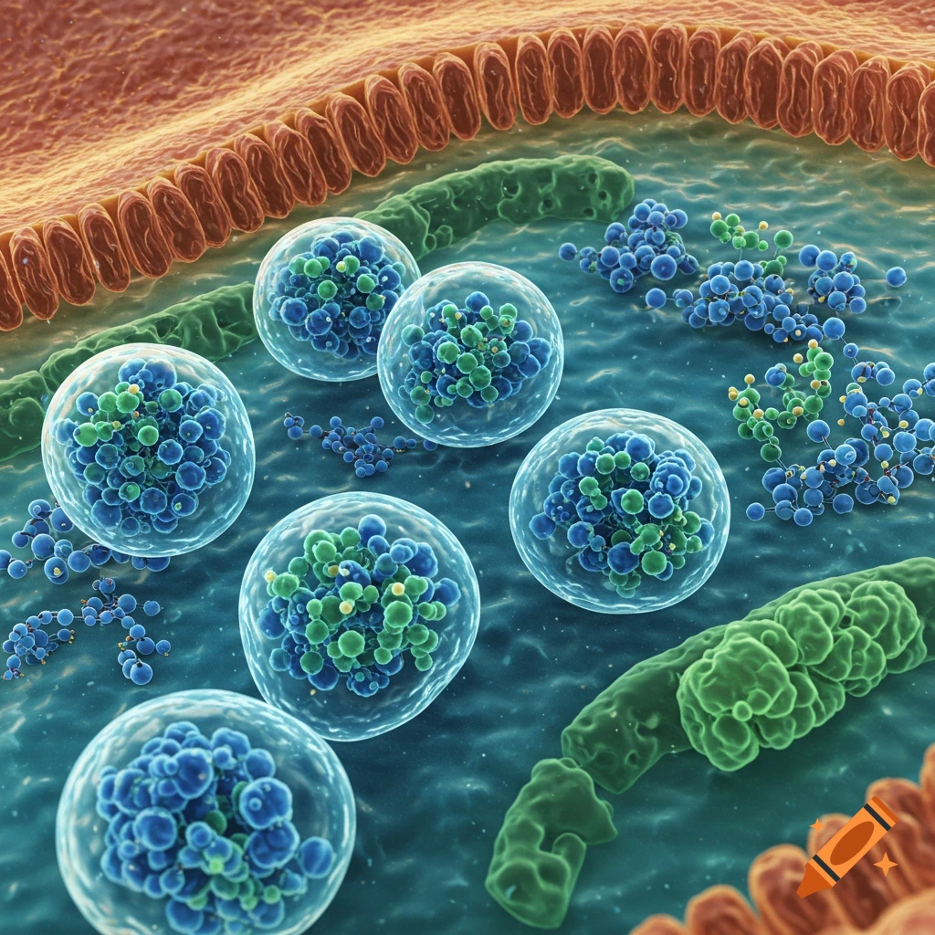
Zoomed-in three-dimensional cross-section of a cell showing biomolecular condensates and cellular structures.
Zoom-Aktion
High-magnification histology slide of ovarian tissue, showing follicles and stromal cells.
Zoom-Aktion
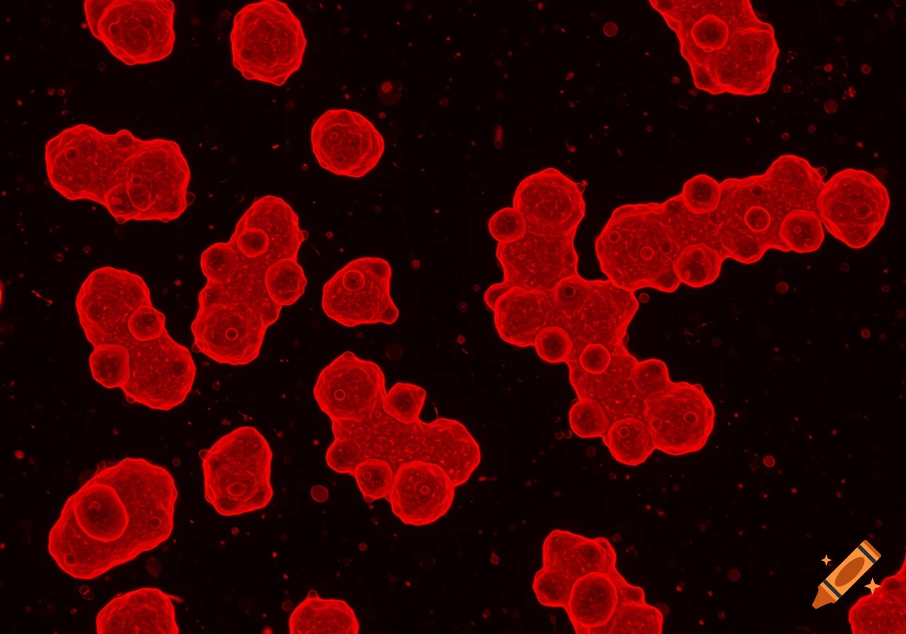
Red virus-like cells viewed through a microscope against a black background.
Zoom-Aktion
Close-up microscopic view of pink fibrous connective tissue against a blue background.
Zoom-Aktion
Microscopic view of bacteria fimbriae
Zoom-Aktion
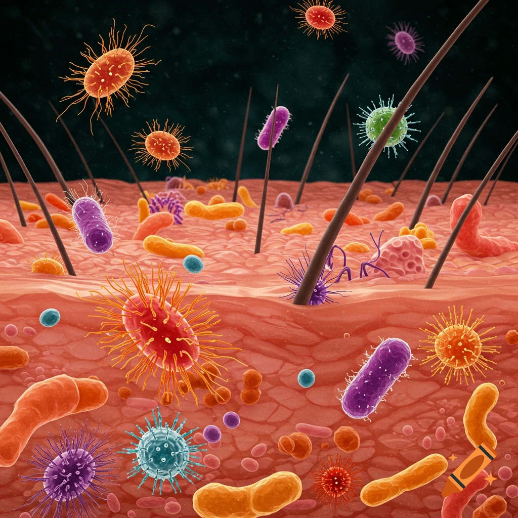
Microscopic view of colorful bacteria and viruses on skin and hair follicles.
Zoom-Aktion
A rotifer under a microscope, swimming among other microorganisms.
Zoom-Aktion
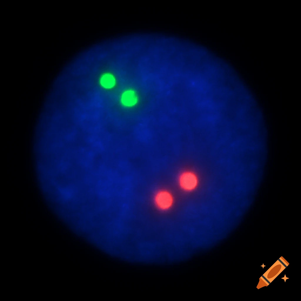
Microscopic view of a cell with glowing green and red spots representing chromosomes.
Zoom-Aktion
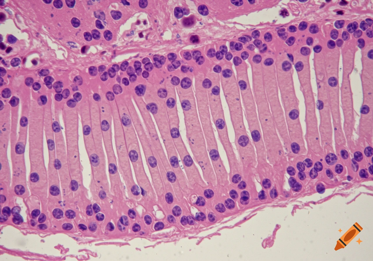
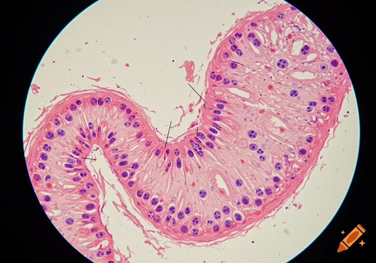
Microscopic view of stomach tissue with pink and purple cells
Zoom-Aktion
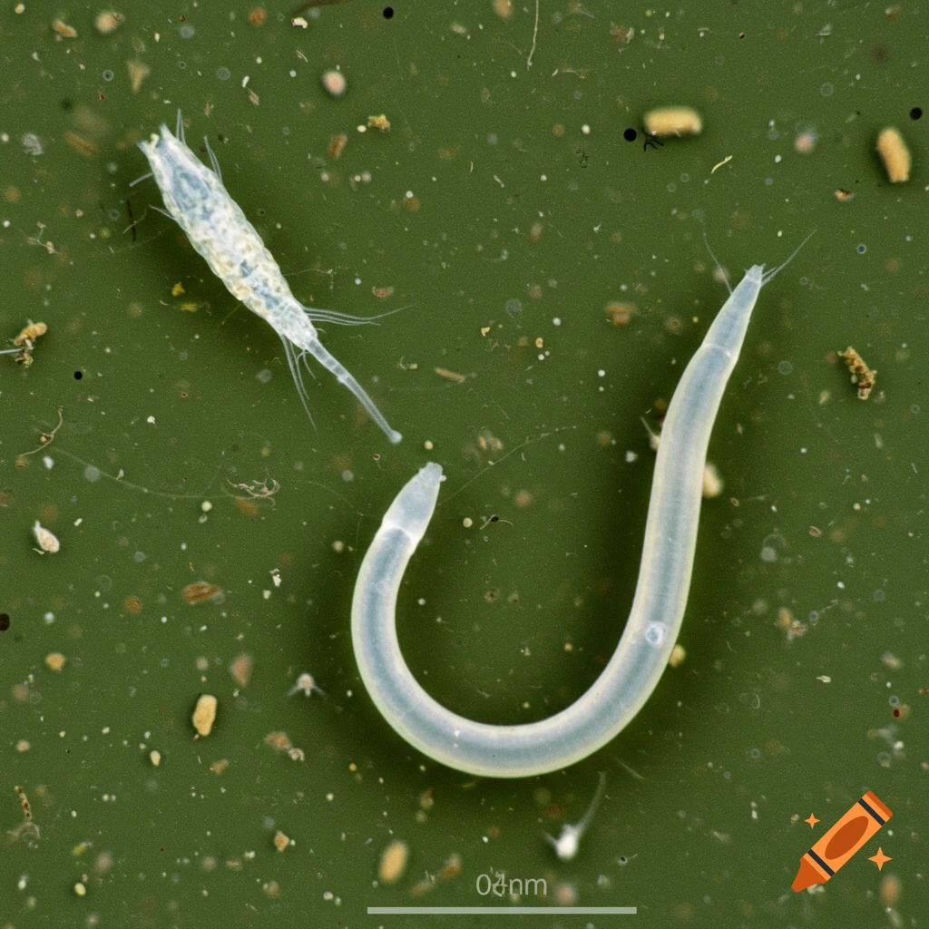
Microscopic view of a white worm and a crustacean-like organism with a scale bar.
Zoom-Aktion
Microscopy image showing ceramic grains and grain boundaries
Zoom-Aktion
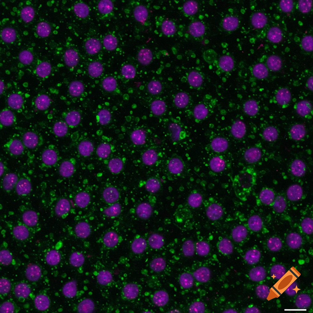
Fluorescence microscopy image showing purple cell nuclei surrounded by green cytoplasm on a dark background.
Zoom-Aktion
Fluorescent microscope image of cells with blue nuclei and red structures on a black background.
Zoom-Aktion
Microscopic view of cells with blue nuclei and red filamentous structures on a black background.
Zoom-Aktion
Immunofluorescence images of GAP-43 staining showing axonal regrowth post-tumor resection
Zoom-Aktion
Microscopic view of tiny person in large uterine cavity
Zoom-Aktion
Bromine-Yeast Oxide viewed under microscope
Zoom-Aktion
Chromatographic resin beads viewed under a microscope
Zoom-Aktion
Ameba under a microscope showing its irregular shape and internal structures like the nucleus and pseudopods
Zoom-Aktion
Genetics and electron microscope in high definition
Zoom-Aktion
Genetics and electron microscope in high definition
Zoom-Aktion
Genetics and electron microscope in high definition
Zoom-Aktion
Microscope images of animal cells on white background
Zoom-Aktion
Microscopic view of cordyceps infection
Zoom-Aktion
Blood drop with microscope inside
Zoom-Aktion
Fluorescent gene transcript in cells of tissue in 3D
Zoom-Aktion
Close-up view of leaf veins under an electron microscope
Zoom-Aktion
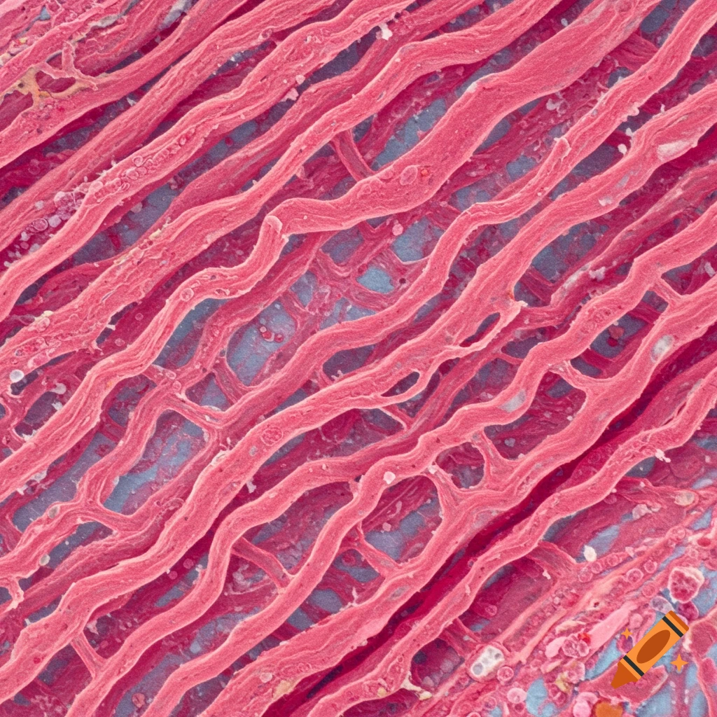
Intricately-detailed muscle fibers under a microscope
Zoom-Aktion
Extracellular vesicle
Zoom-Aktion
Microscopic view of a mutant diatom
Zoom-Aktion
High magnification of translucent membrane with histological stains
Zoom-Aktion
SEM image of cobalt oxide
Zoom-Aktion
3D morphology of nickel foam coated with Carbon Nanotube resembling coral growth
Zoom-Aktion
Microscopic view
Zoom-Aktion
Microscopic view
Zoom-Aktion
Genetics and electron microscopy exploring the intricate tapestry of life at a hyperdetailed level with 3D renders
Zoom-Aktion
Genetics and electron microscopy exploring the intricate tapestry of life at a hyperdetailed level with 3D renders
Zoom-Aktion
Genetics and electron microscopy exploring the intricate tapestry of life at a hyperdetailed level with 3D renders
Zoom-Aktion
Stained cells for specific proteins or markers
Zoom-Aktion
Microscopic view of a brain
Zoom-Aktion
high contrast seamless pattern of human lung cells
Zoom-Aktion
colony of yeast cells on an agar plate
Zoom-Aktion
scientist examining viruses under a microscope
Zoom-Aktion
microscopy image for product marketing management
Zoom-Aktion
microscope images of amoebae
Zoom-Aktion
image of an artificial cell
Zoom-Aktion
vibrant microscopy image of cyanobacterial filaments
Zoom-Aktion
fractal fungal growth inspired by Haeckel
Zoom-Aktion
image of a compound microscope
Zoom-Aktion
visualization of a cell nucleus
Zoom-Aktion
microscopic view of an ibroblast
Zoom-Aktion
microscopic view of Sapphire Blue Metallic
Zoom-Aktion
microscopic view of a crystal diamond texture
Zoom-Aktion
fluorescent microscopy image of bacteria
Zoom-Aktion
microscopic view of cancer cells
Zoom-Aktion
close-up view of bacterial fibers capturing bacteria
Zoom-Aktion
black and white scanning electron microscopy of a goat willow flower
Zoom-Aktion
black and white microscopy of a rhododendron flower
Zoom-Aktion
black and white microscopy of a buddleja flower
Zoom-Aktion
black and white scanning electron microscopy of leather
Zoom-Aktion
scanning electron microscopy of lichen cells
Zoom-Aktion
black and white SEM image of borage
Zoom-Aktion
black and white microscopy image of lichen
Zoom-Aktion
black and white image of flower petals under electron microscope
Zoom-Aktion
black and white microscopic image of flower pollen
Zoom-Aktion
microscopic image of aligned polymeric nanofibers
Zoom-Aktion
visual representation of cell-cell interaction
Zoom-Aktion
microscopic view of human esophagus cells
Zoom-Aktion
microscopic view of a blood smear
Zoom-Aktion
microscopy image of human pancreatic cells
Zoom-Aktion
microscopic view of a human hair cell
Zoom-Aktion
microscopic view of human blood cells
Zoom-Aktion
microscopic view of human lung cells
Zoom-Aktion
microscopic view of human liver cells
Zoom-Aktion
microscopic view of a human nerve cell
Zoom-Aktion
detalle microscópico de células del corazón humano
Zoom-Aktion
microscopic view of a cat, diatoms and bacteria
Zoom-Aktion
microscopic image of a bumpy animal cell
Zoom-Aktion
ultrastructure microscopic image
Zoom-Aktion