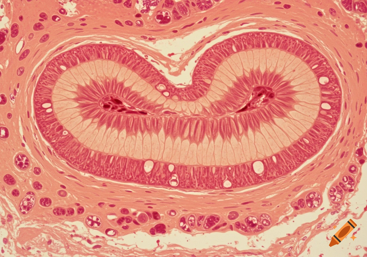
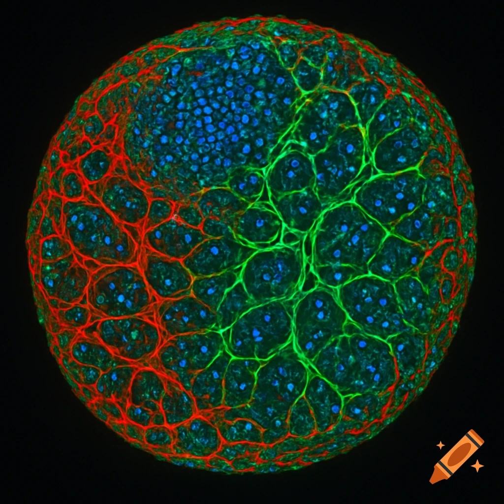
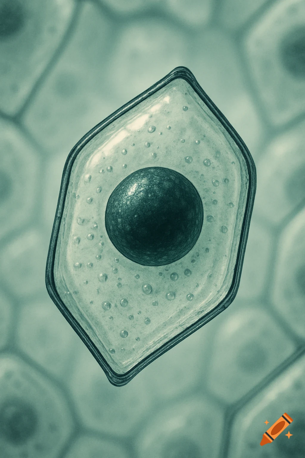
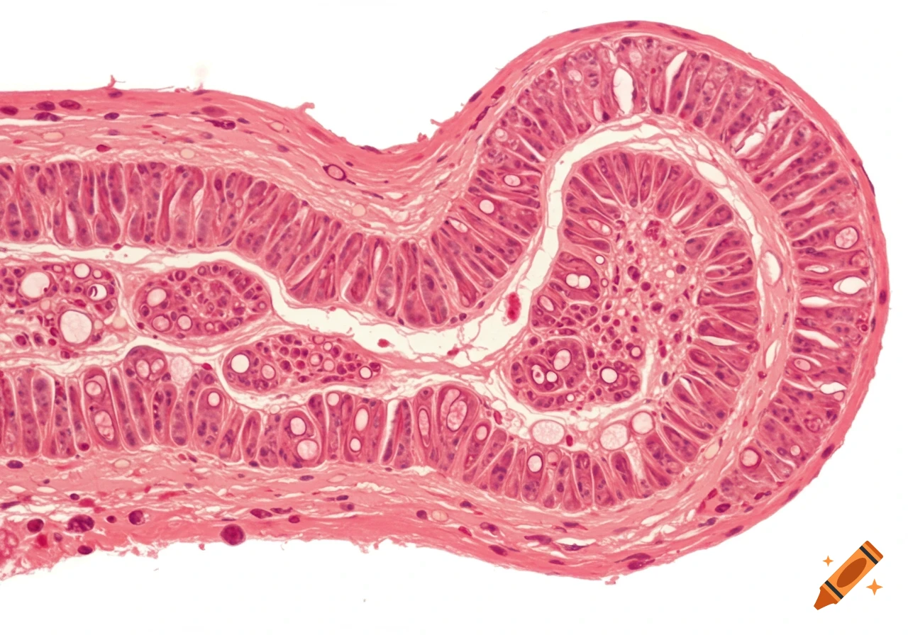
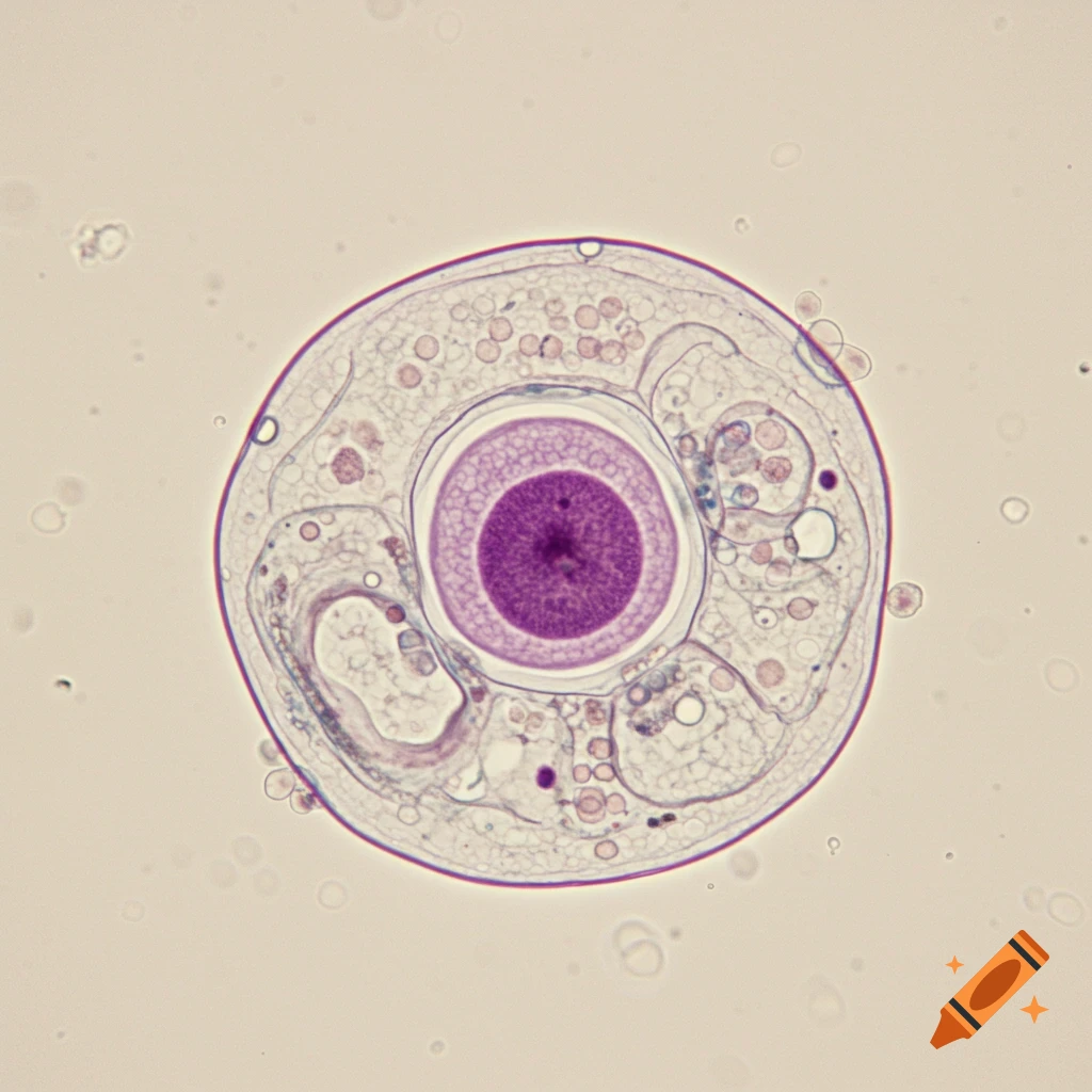
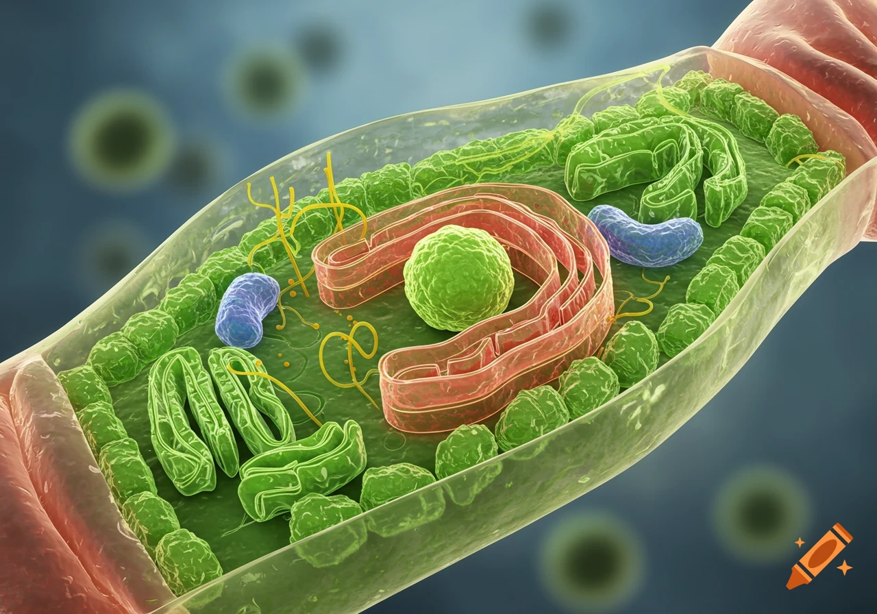
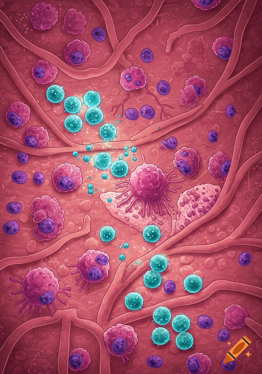
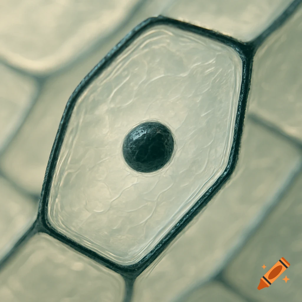
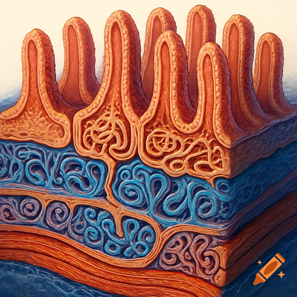
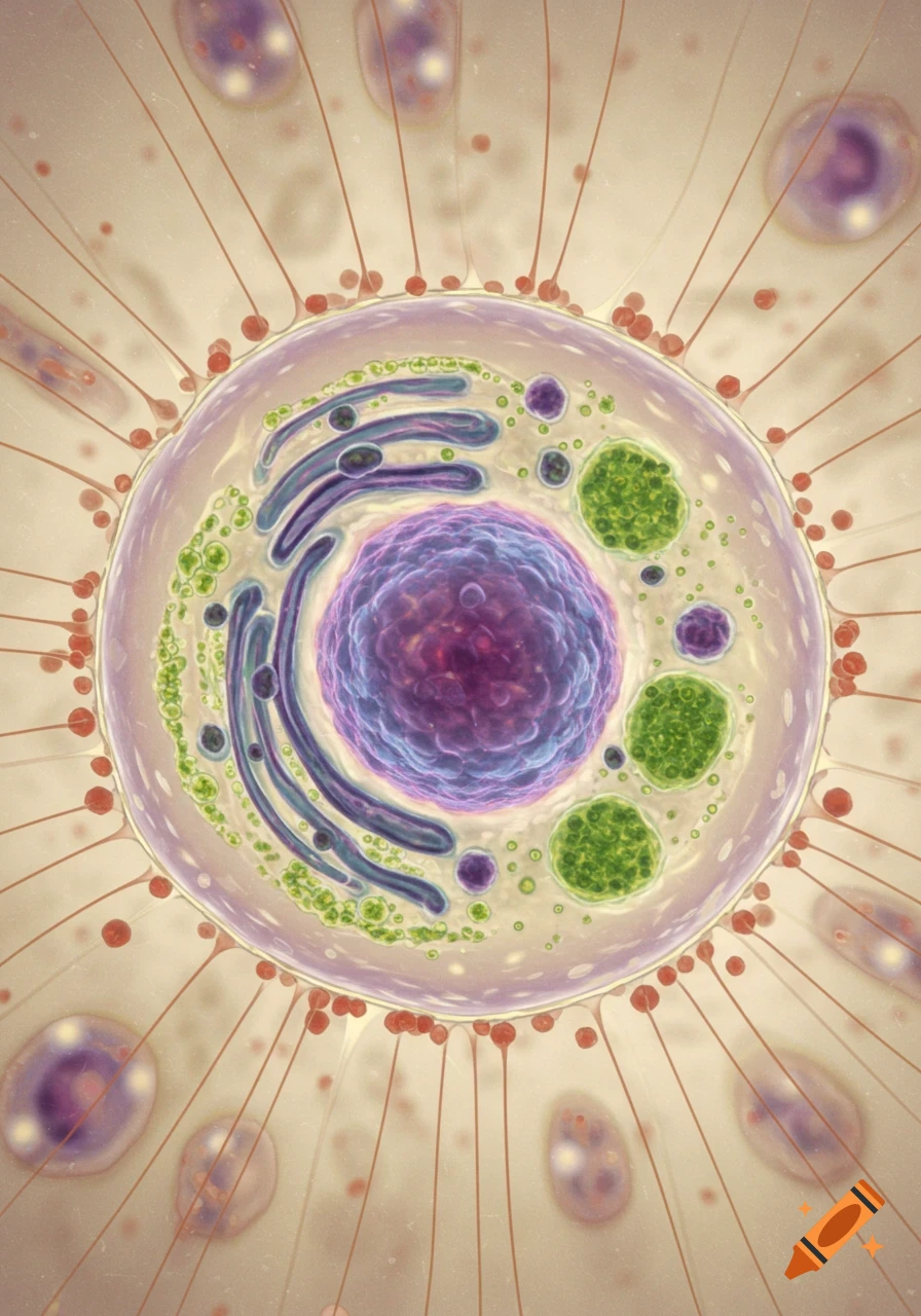
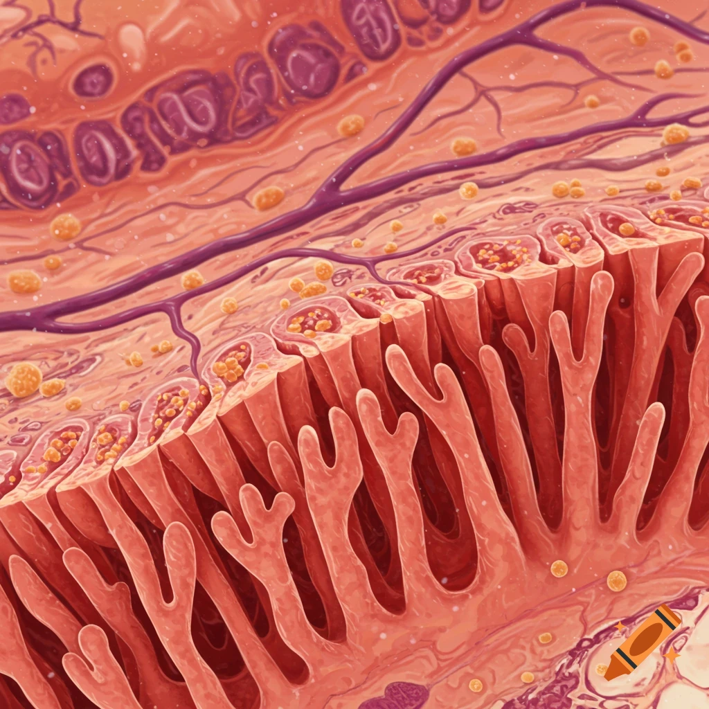
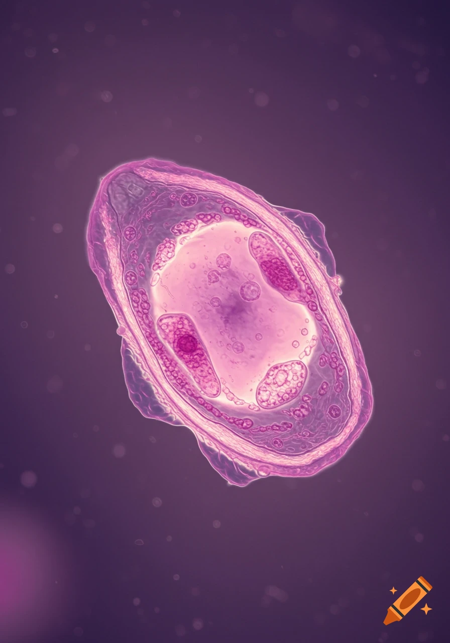
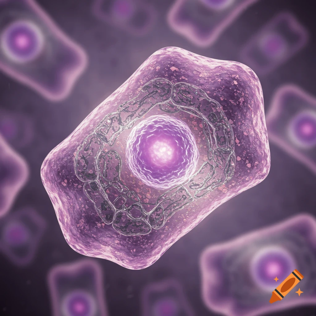
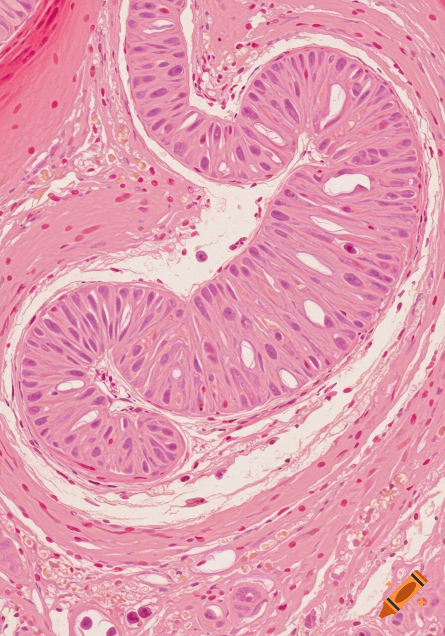
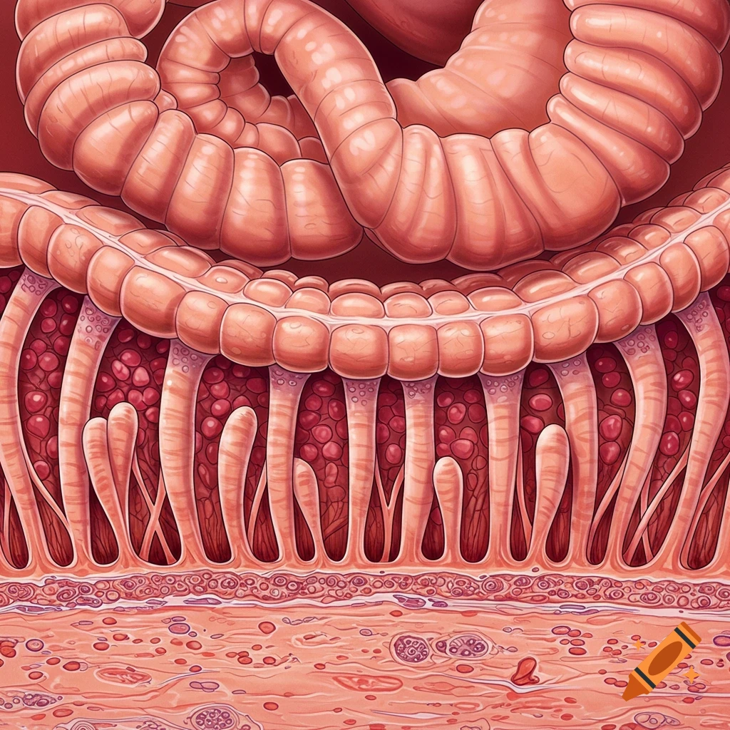
Close-up microscopic view of simple columnar epithelium tissue, stained pink and purple, showing elongated cells and visible nuclei.
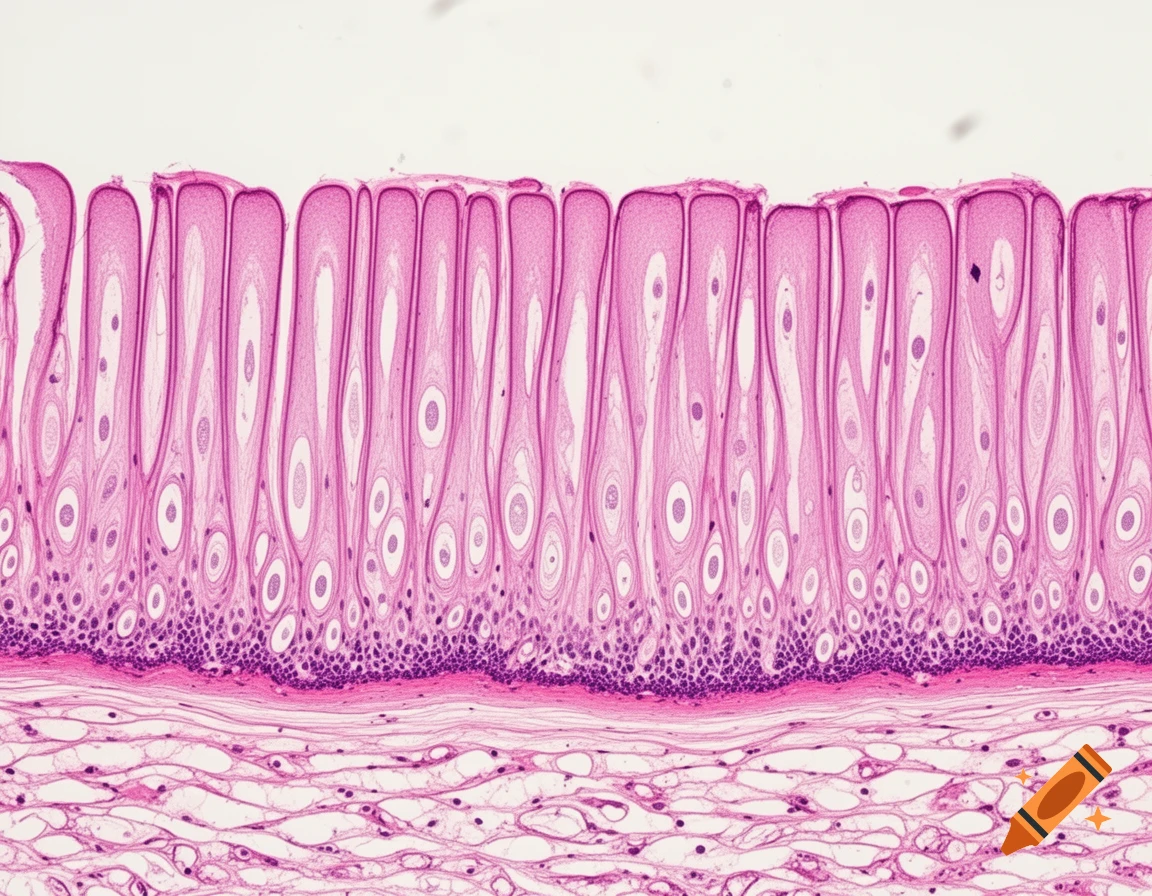
A highly realistic microscopic image of simple columnar epithelium tissue as seen under a standard light microscope. The tissue shows a single layer of tall, closely packed columnar cells with elongated, oval nuclei aligned near the basal region. Each cell contains clearly visible cytoplasm with soft pink staining. A distinct basement membrane is present at the base of the epithelial layer, separating it from underlying connective tissue. Several goblet cells are interspersed among the columnar cells, appearing pale and mucus-filled with compressed nuclei. The image resembles an H&E-stained histology slide, high magnification, sharp cellular detail, realistic medical laboratory appearance, educational anatomy style. Mehr sehen
More images like this