A scientific diagram illustrating the V2-SPG Neuro-Glial Hypothesis, showing a Trigeminal Ganglion and a Sphenopalatine Ganglion with labeled components and paracrine glial cross-talk.
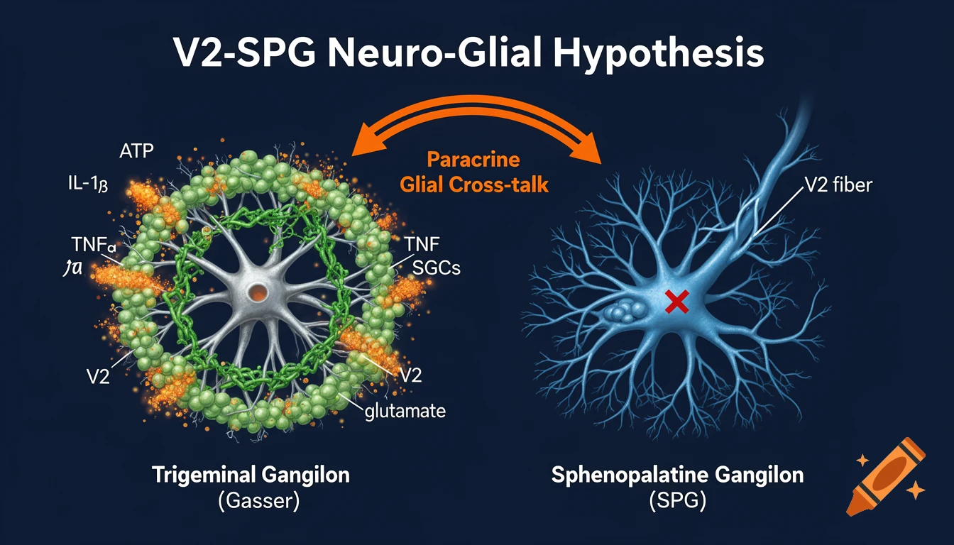
Scientific medical infographic diagram, dark navy blue background, style of Nature Reviews Neuroscience. Shows two ganglia side by side. LEFT: Trigeminal Ganglion (Gasser) with large central neuron (V2 maxillary branch) completely surrounded by satellite glial cells (SGCs) colored in green. Glial cells releasing orange labeled particles: ATP, IL-1beta, TNF-alpha, glutamate. Green gap junctions connecting glial cells. RIGHT: Sphenopalatine Ganglion (SPG) with parasympathetic neurons in blue. V2 fiber shown passing through SPG with a red X symbol indicating NO direct synapse. Orange curved double arrow between the two ganglia labeled Paracrine Glial Cross-talk. Bold white title at top: V2-SPG Neuro-Glial Hypothesis. Clean white scientific labels. High resolution professional medical illustration Ver mais
More images like this