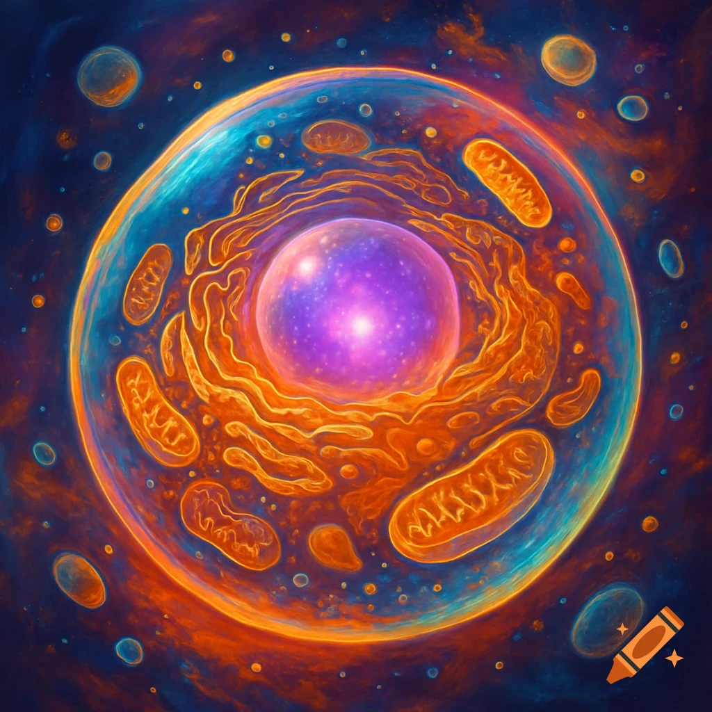
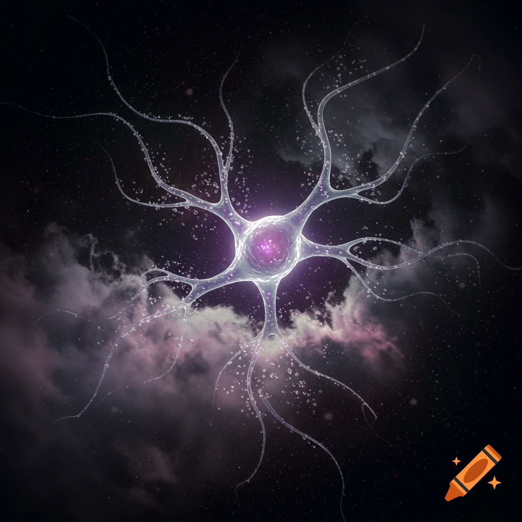
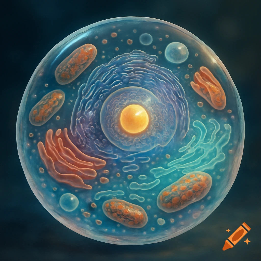
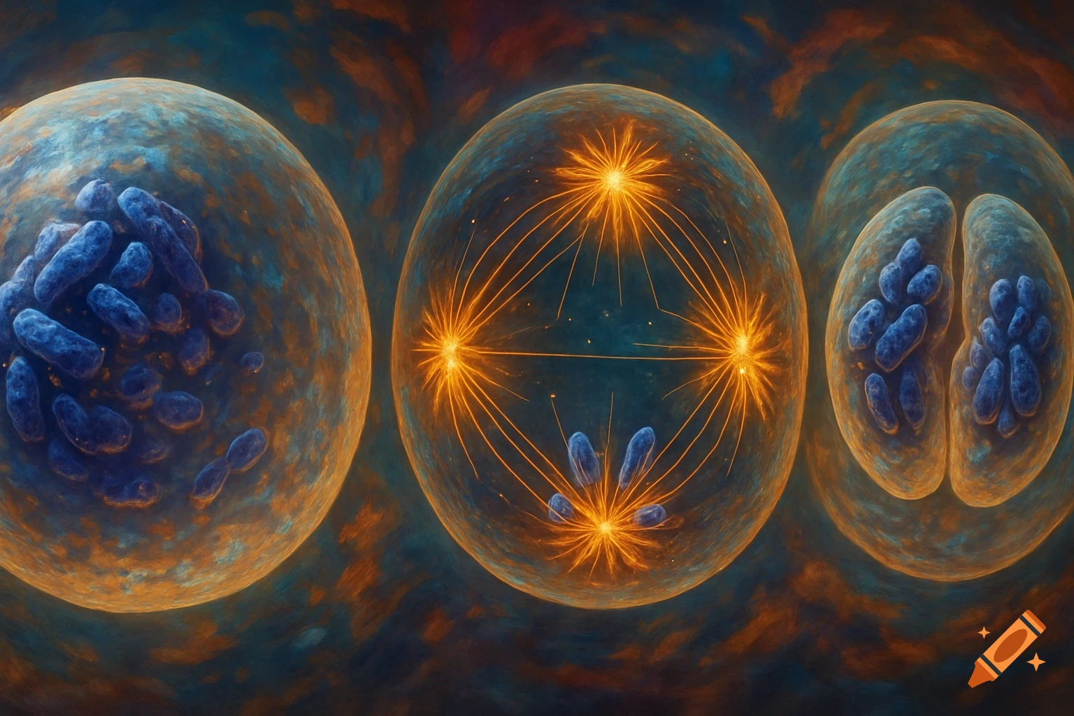
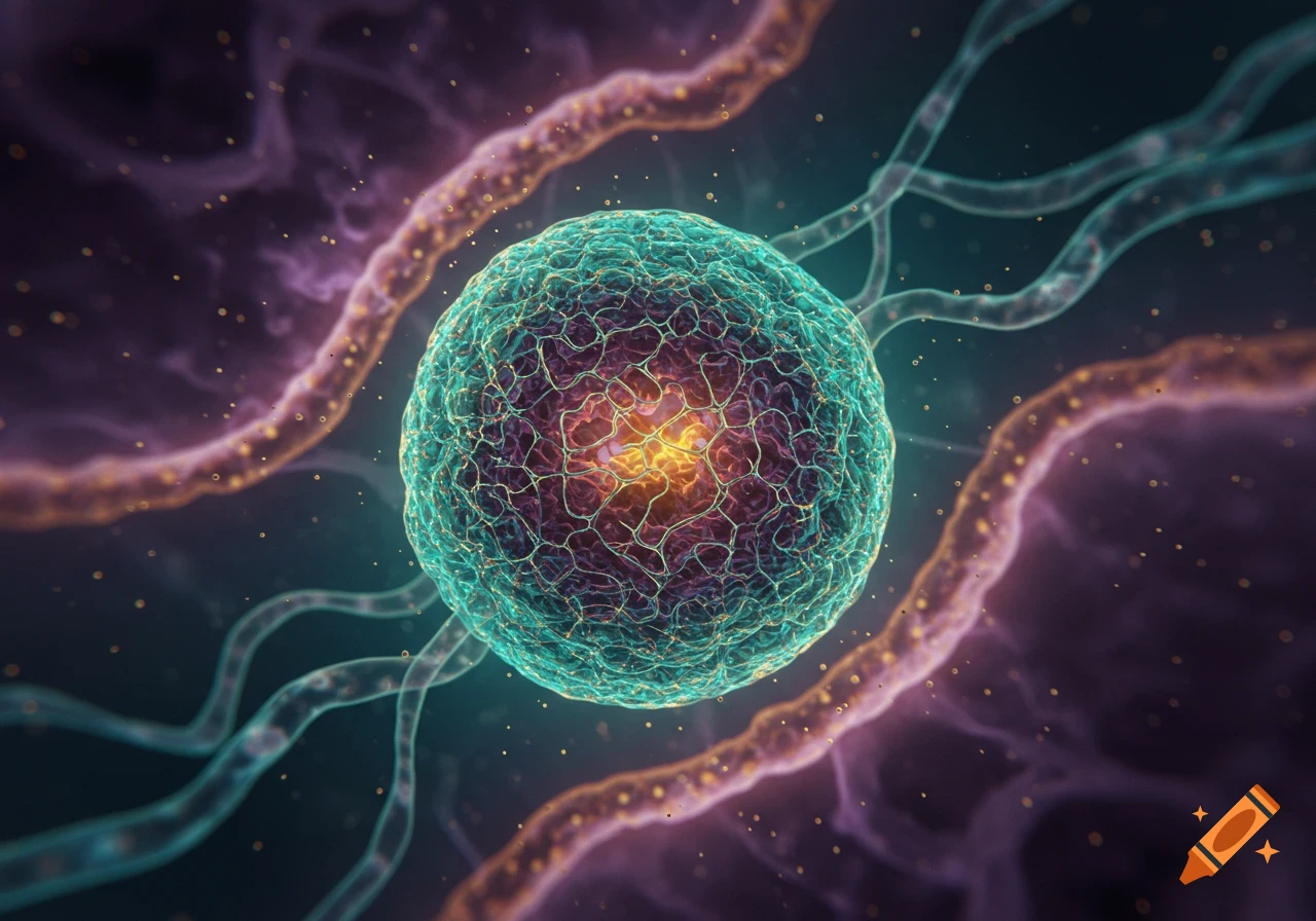
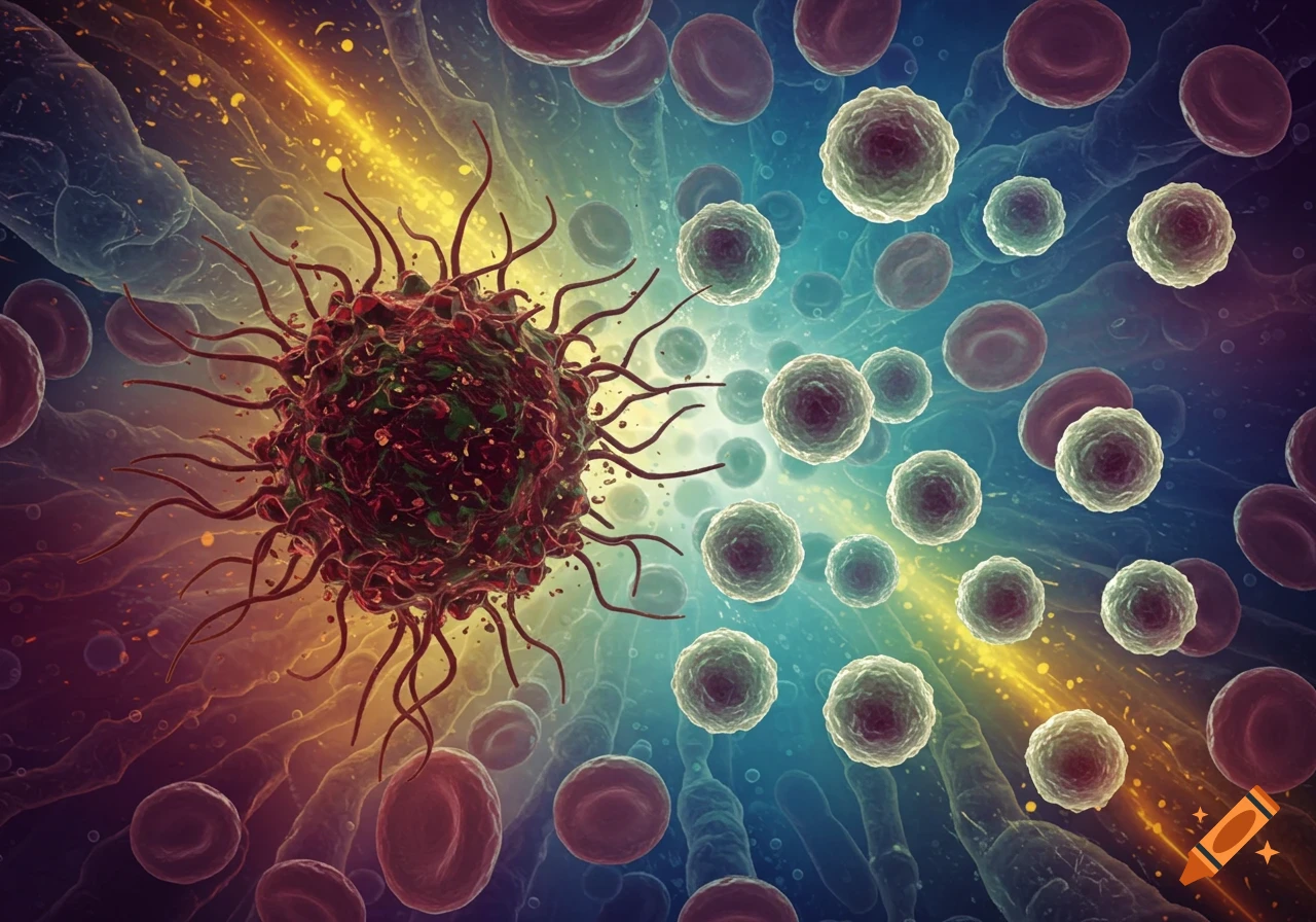
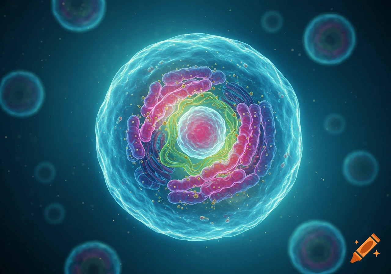
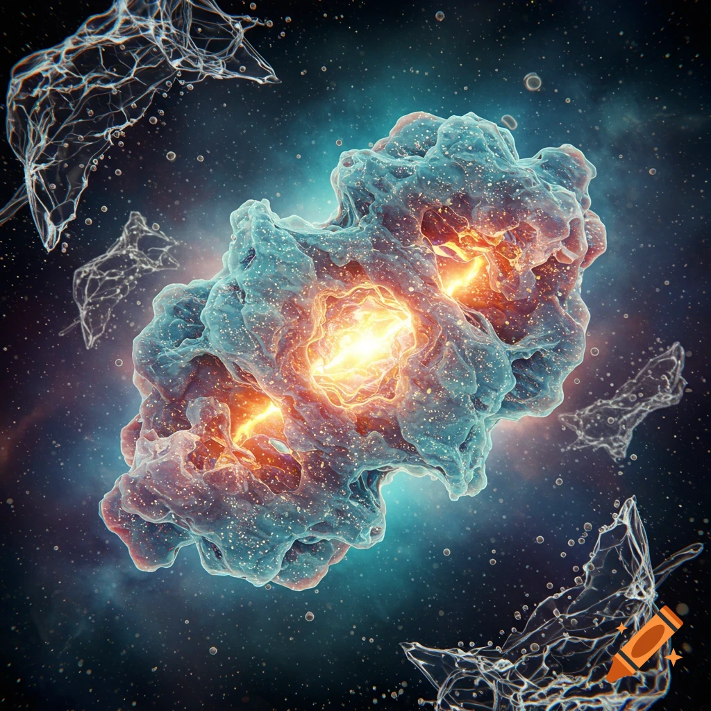
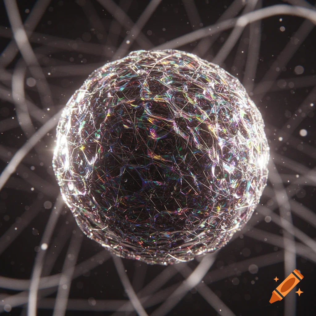
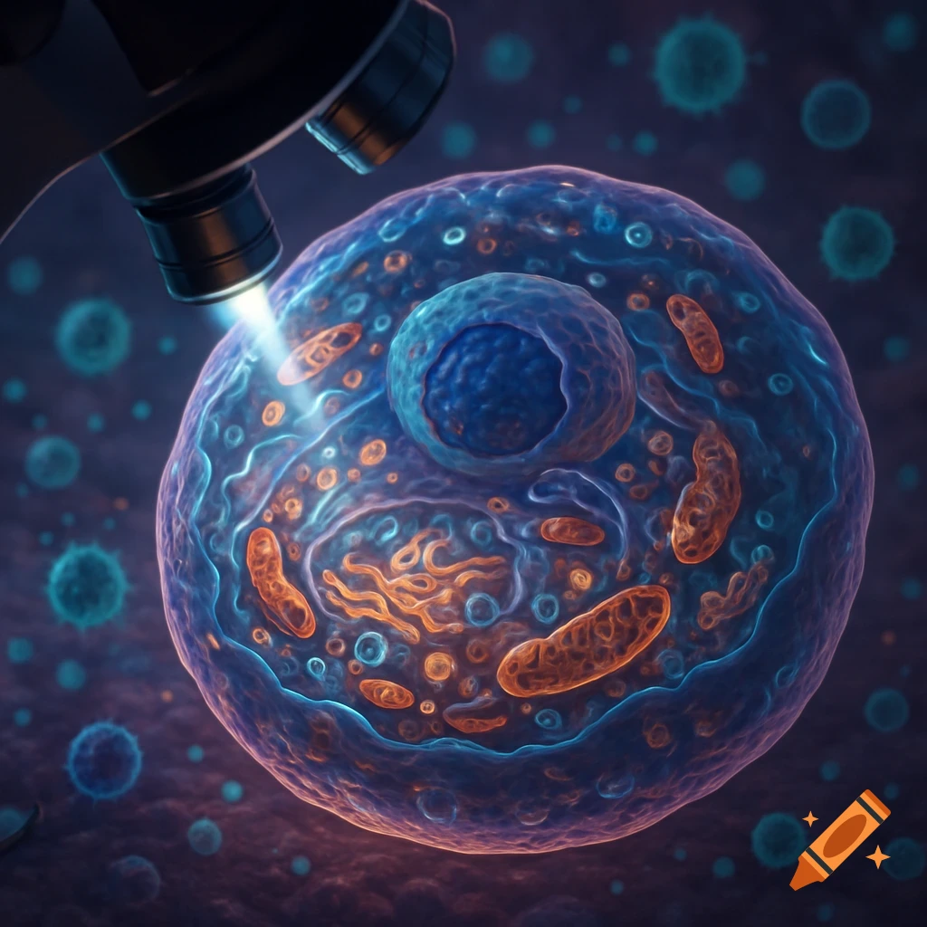
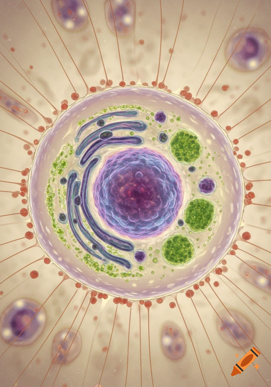
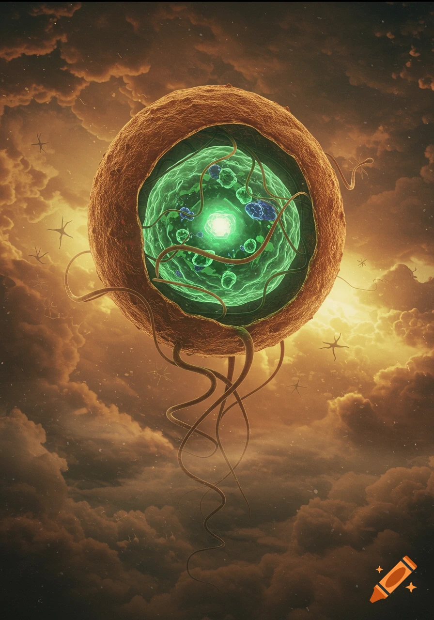
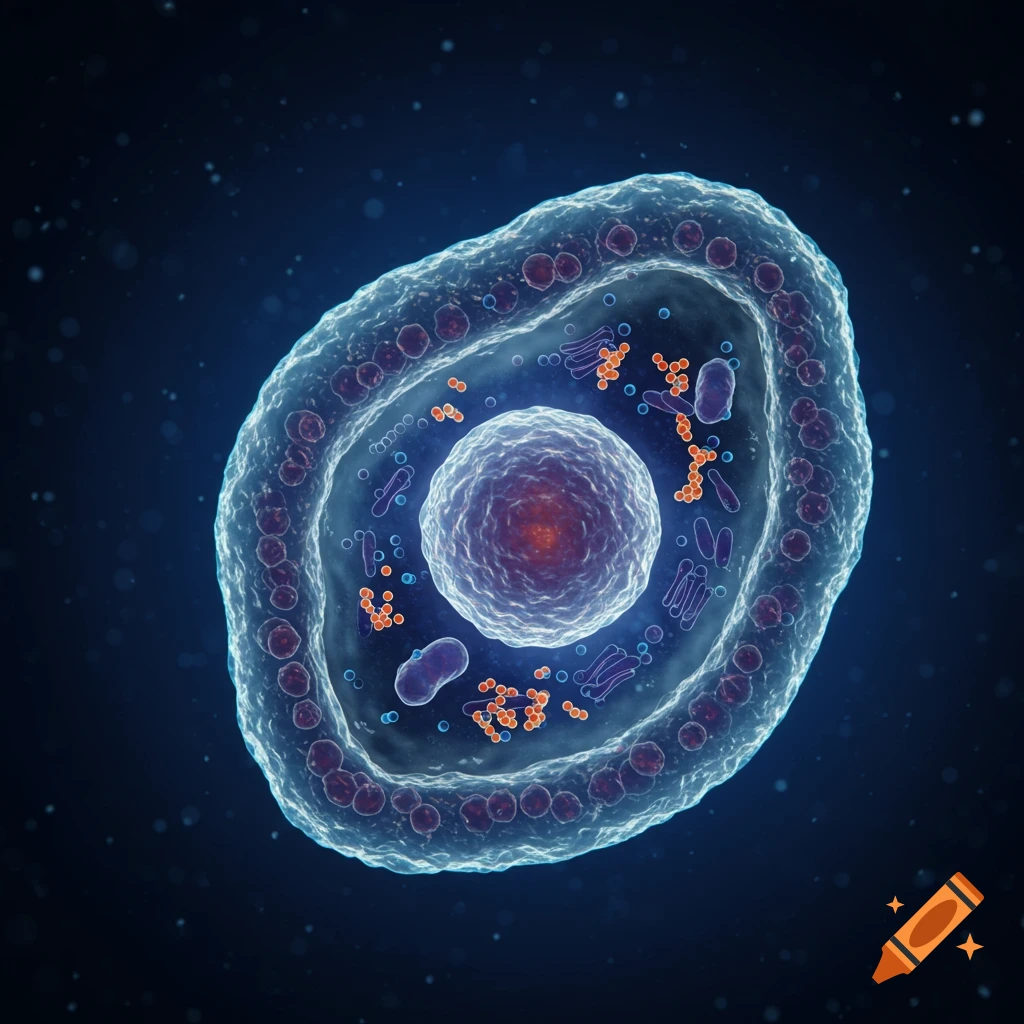
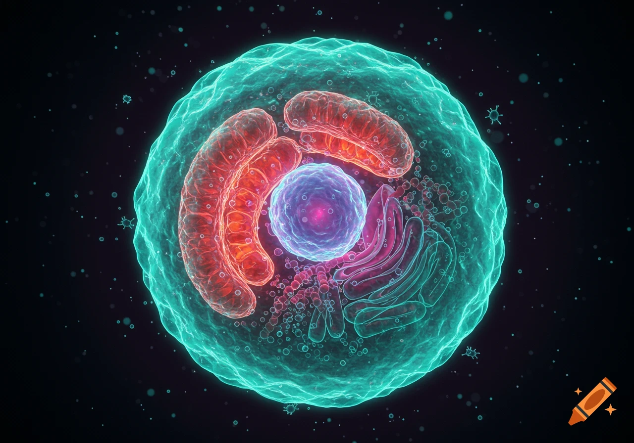
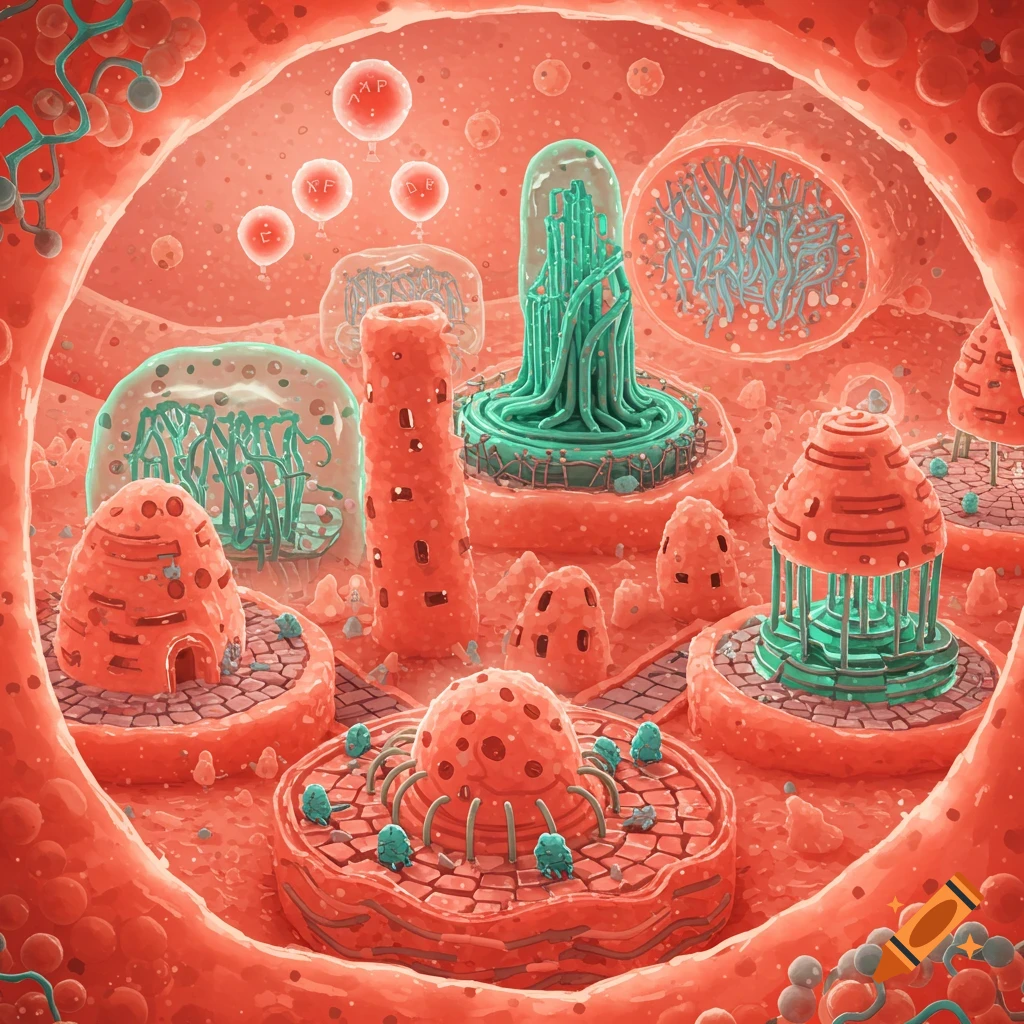
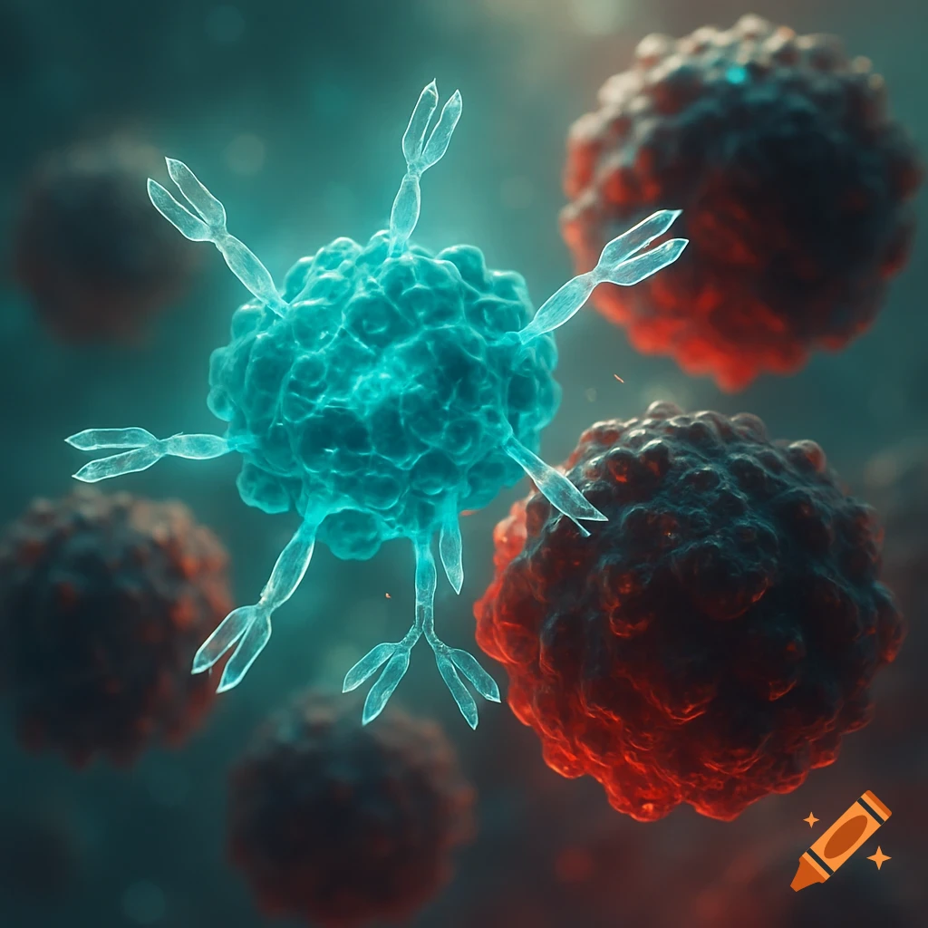
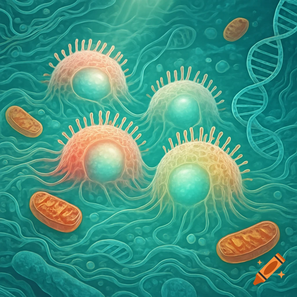
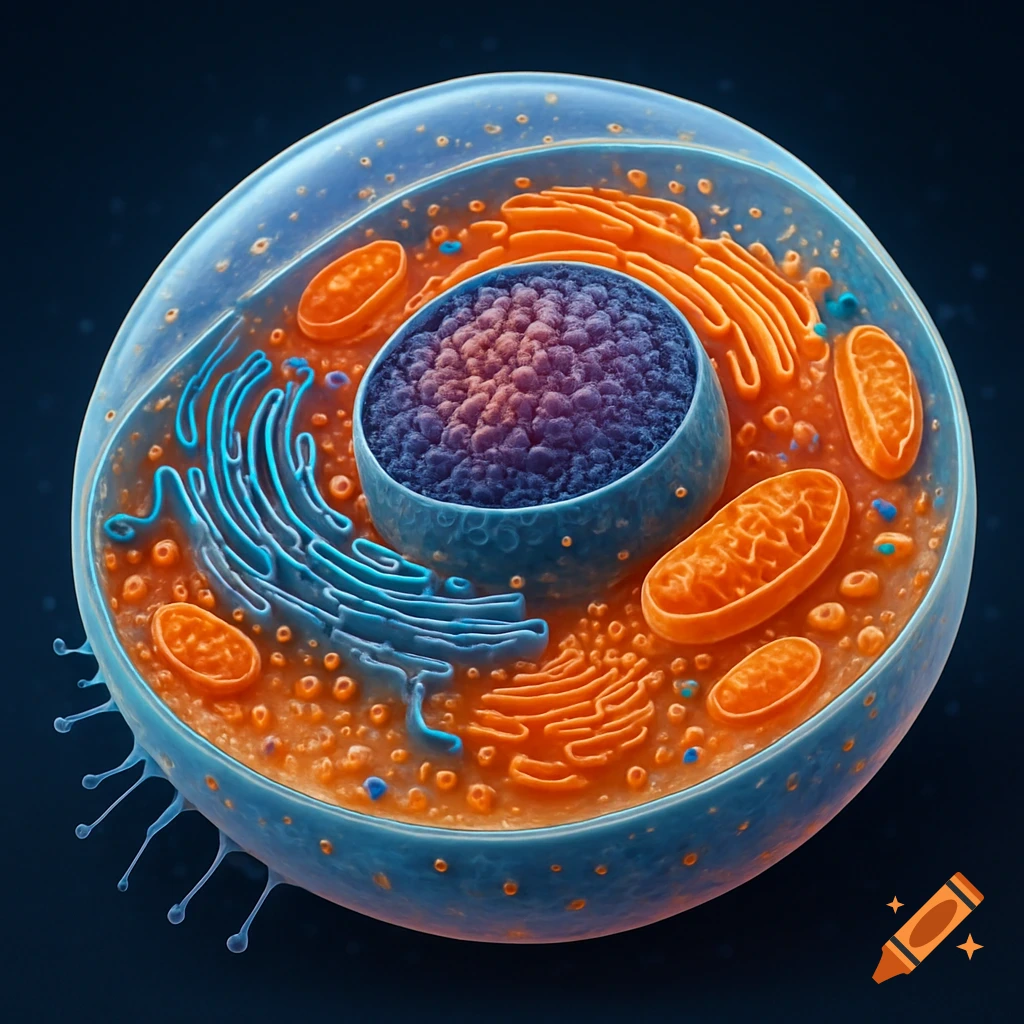
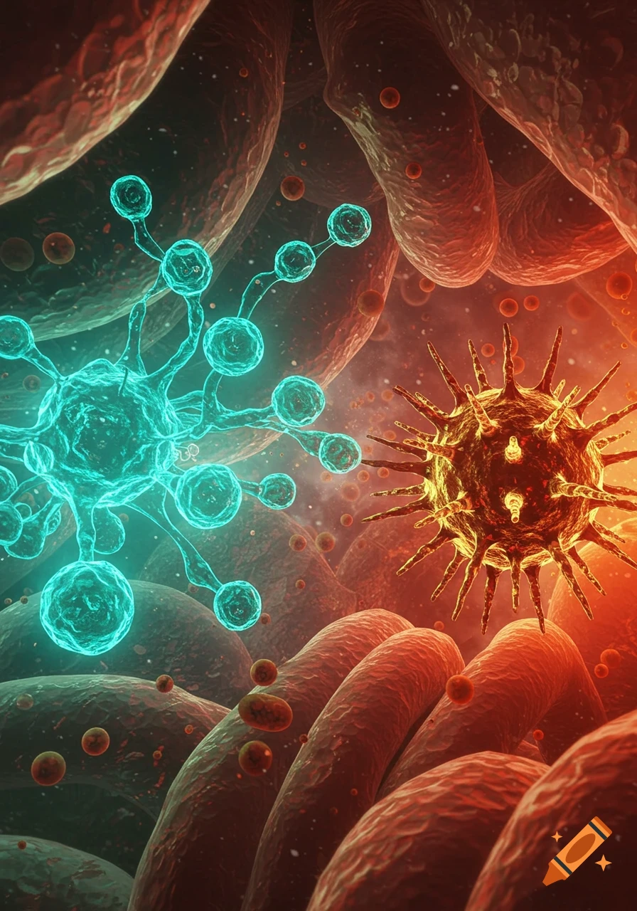
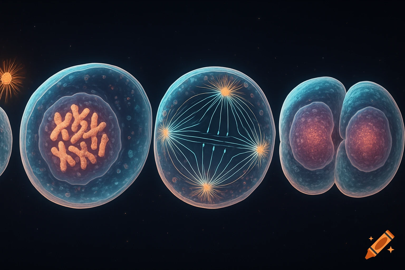
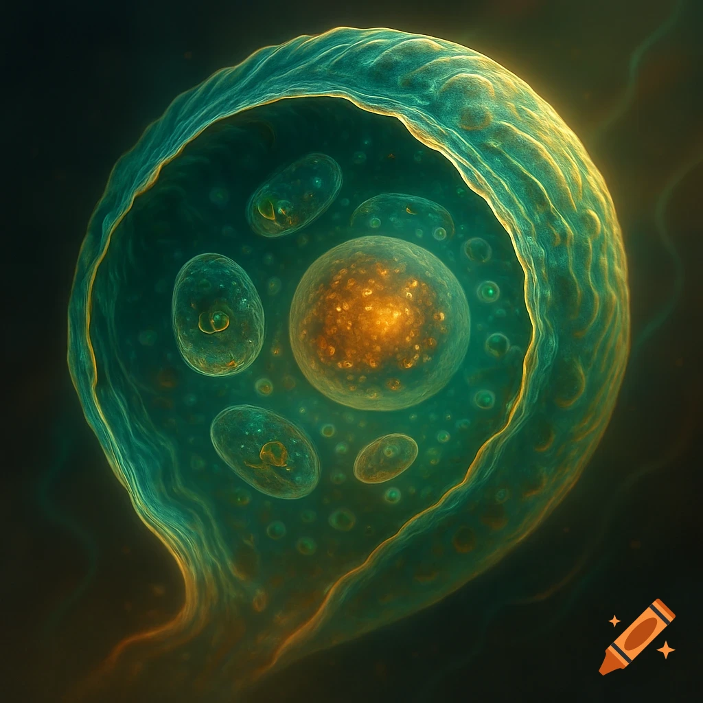
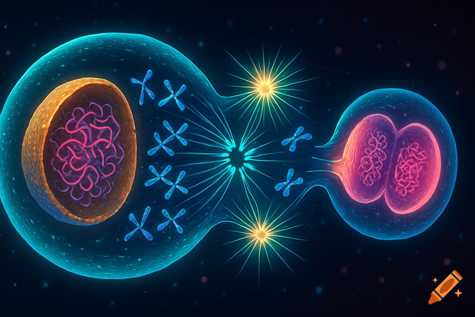
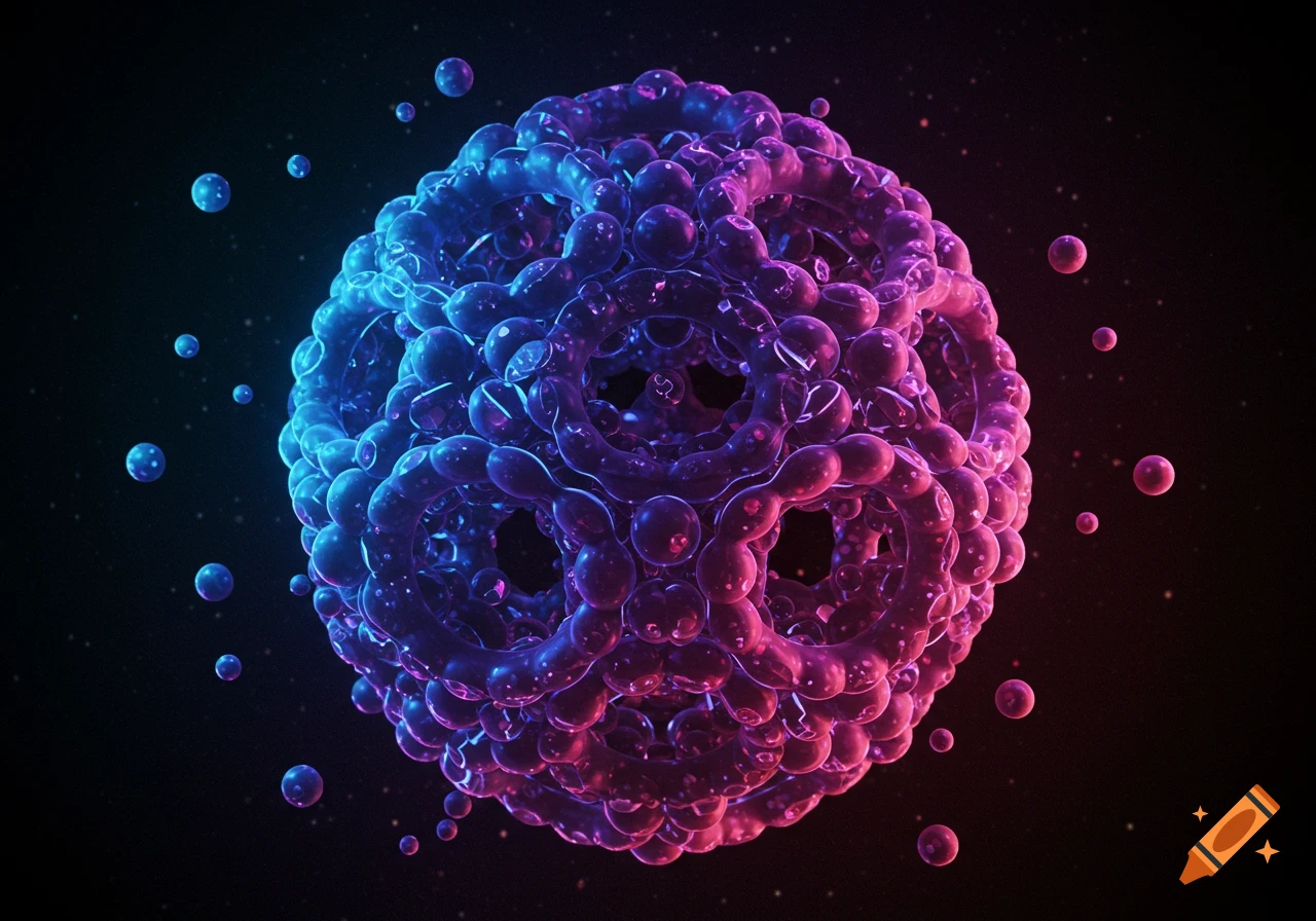
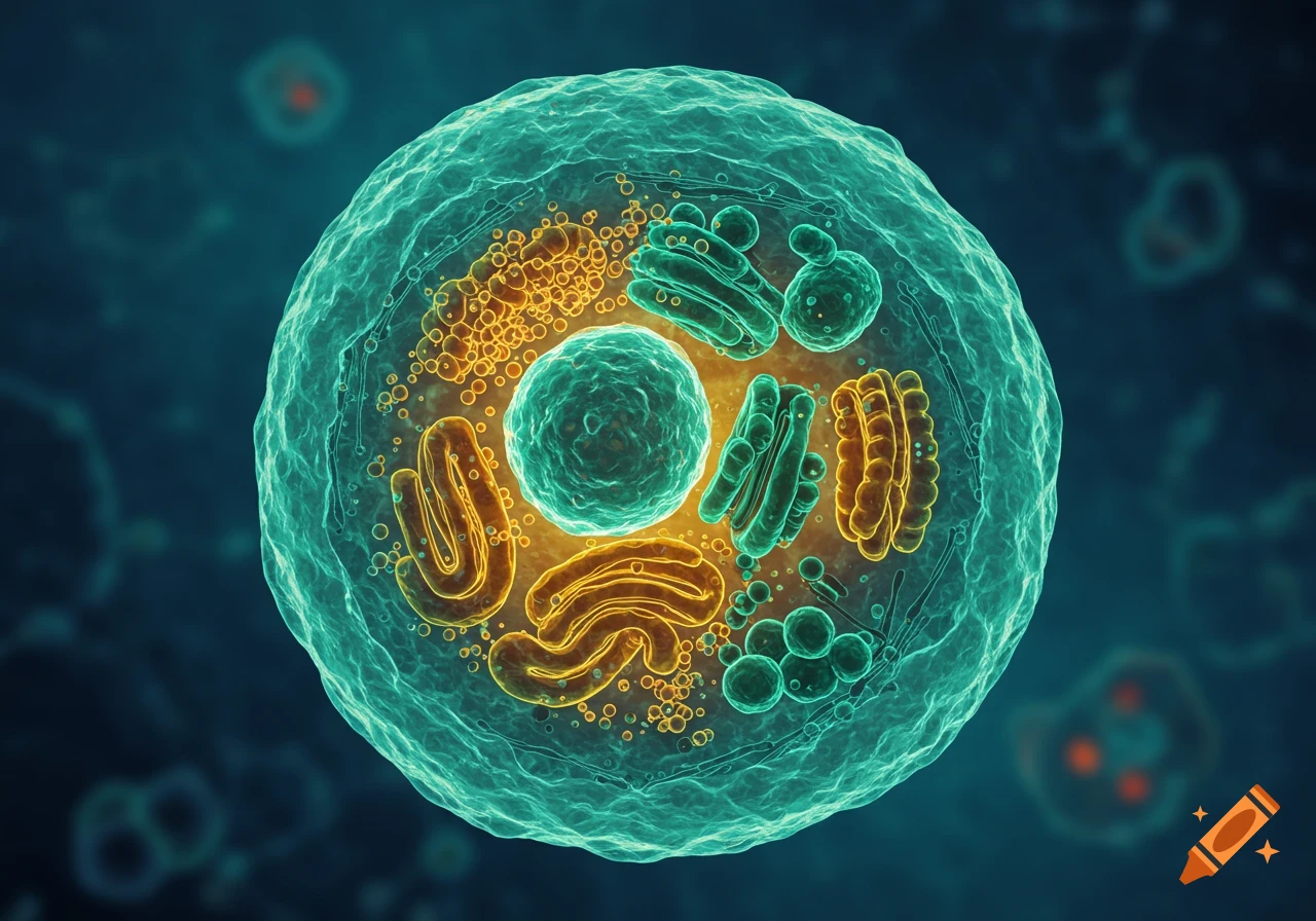
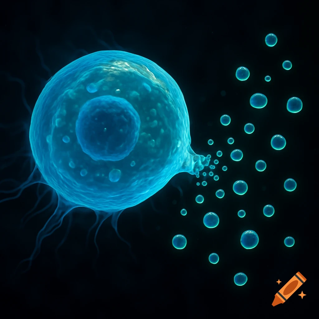
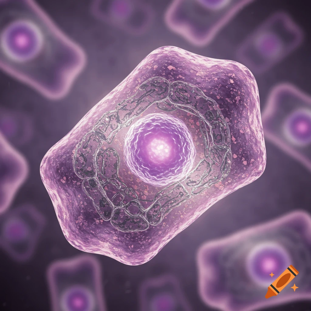
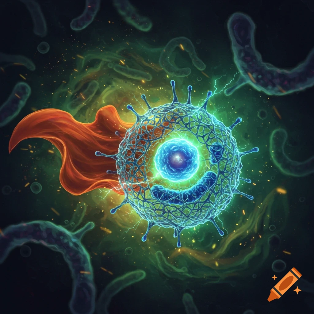
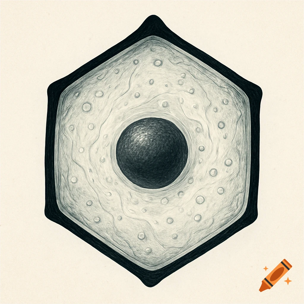
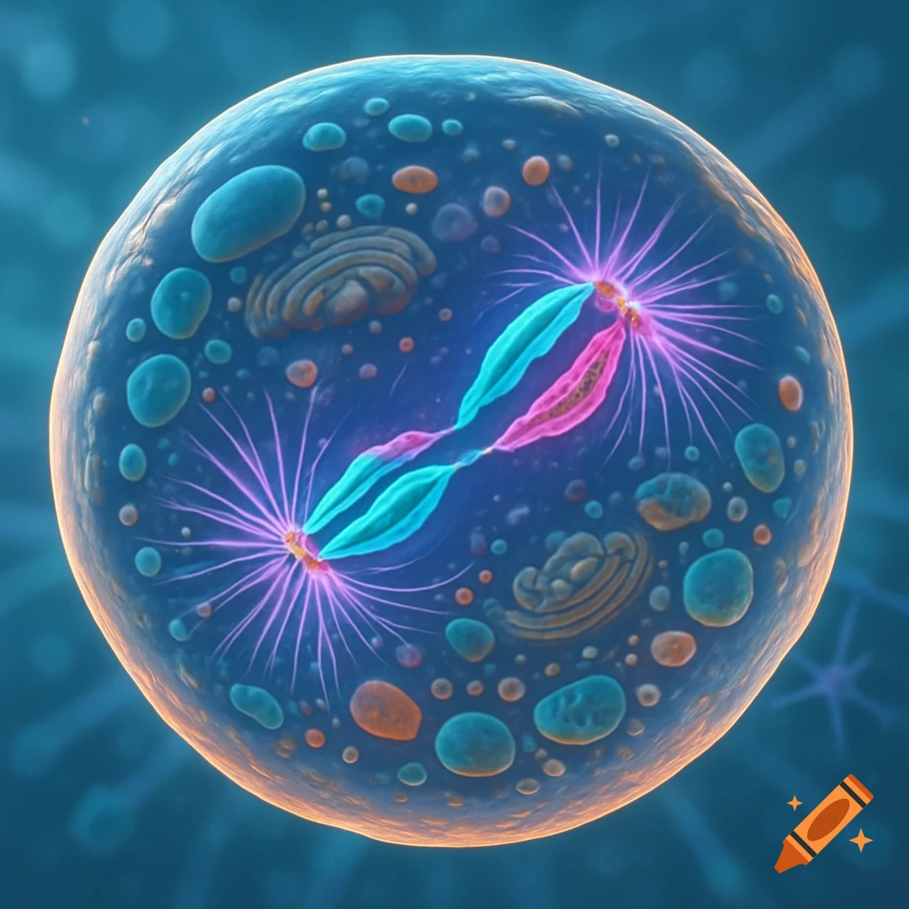
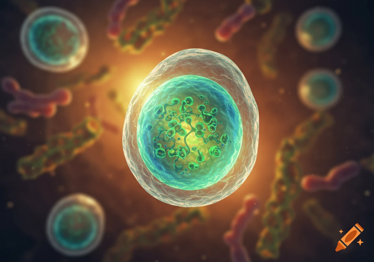
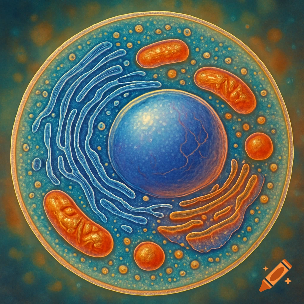
Hyper-realistic scientific illustration of a cell with a central nucleus and many translucent virus particles, on a cracked organic background.

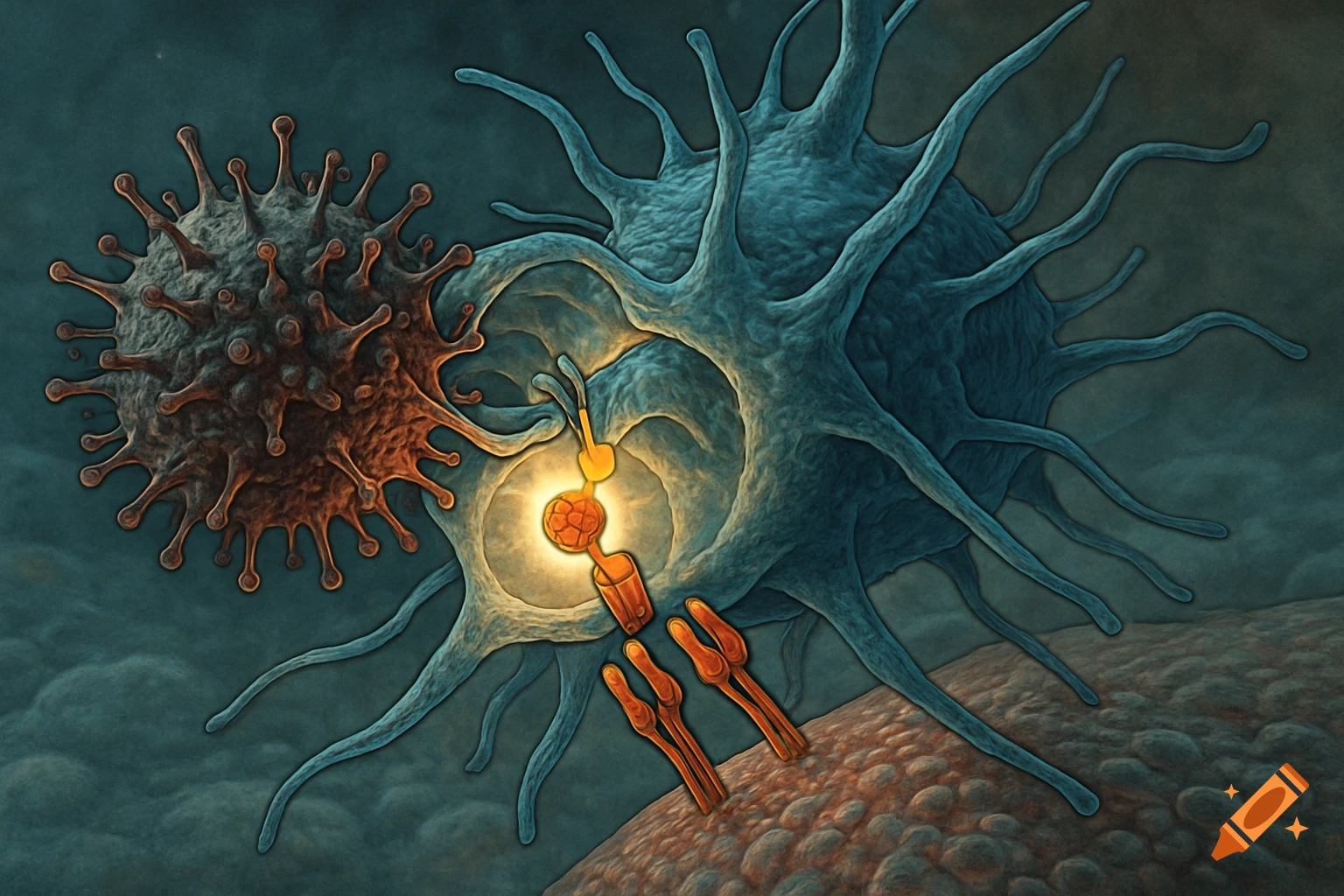
Hyper-realistic scientific illustration of a Lassa virus-infected dendritic cell within a lymph node, rendered in the style of a museum-quality natural history illustration. The dendritic cell is depicted with detailed cellular anatomy—visible nucleus, endoplasmic reticulum, mitochondria—in warm amber and tan tones. Spherical Lassa virus particles (110nm, roughly 200-300 of them) cluster around and within the cell, rendered as small translucent spheres with a subtle sheen and geometric detail. The background shows the lymph node microenvironment: surrounding immune cells, collagen fibers, and tissue architecture in muted beige, cream, and soft gray tones. Dramatic directional lighting similar to the reference—warm side-lighting creating dimensional depth and soft shadows on the cracked stone-textured background of the lymph node. Cross-sectional view. Highly detailed, scientifically accurate, museum specimen photography aesthetic. Professional medical illustration quality. See more
More images like this