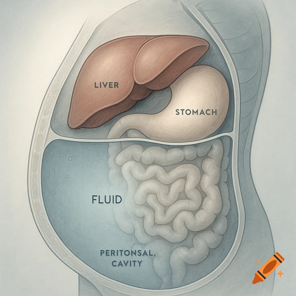
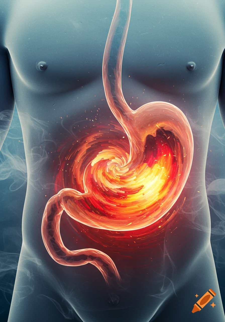
Medical illustration of a Vitzel gastrostomy showing a stomach, feeding tube, sutures, and incision on a human abdomen, with labels.

Medical illustration of Vitzel (Witzel) gastrostomy, side view of human abdomen. Show: 1) Left transrectal laparotomy incision (10–12 cm at costal arch) labeled ‘Incision’, 2) Stomach anterior wall labeled ‘Stomach’, 3) Rubber feeding tube lying on stomach, tip toward pylorus labeled ‘Feeding Tube → Pylorus’, 4) Seroserous canal with 6–8 interrupted serous sutures labeled ‘Seroserous Canal (6–8 Sutures)’, 5) Pyloric region with purse-string suture and 2–3 extra seromuscular sutures labeled ‘Purse-string + 2–3 Seromuscular Sutures’, 6) Tube exiting through separate incision at edge of left rectus muscle labeled ‘Tube Exteriorization’, 7) Gastropexy attaching stomach to parietal peritoneum labeled ‘Gastropexy Sutures’. Educational, anatomical, step-by-step surgical diagram, colored: red = stomach, blue = sutures, green = tube, brown = abdominal wall. Clear labels for each structure.” Mehr sehen
More images like this