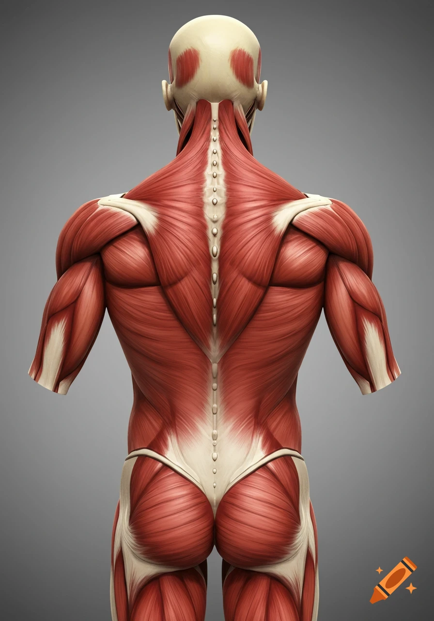
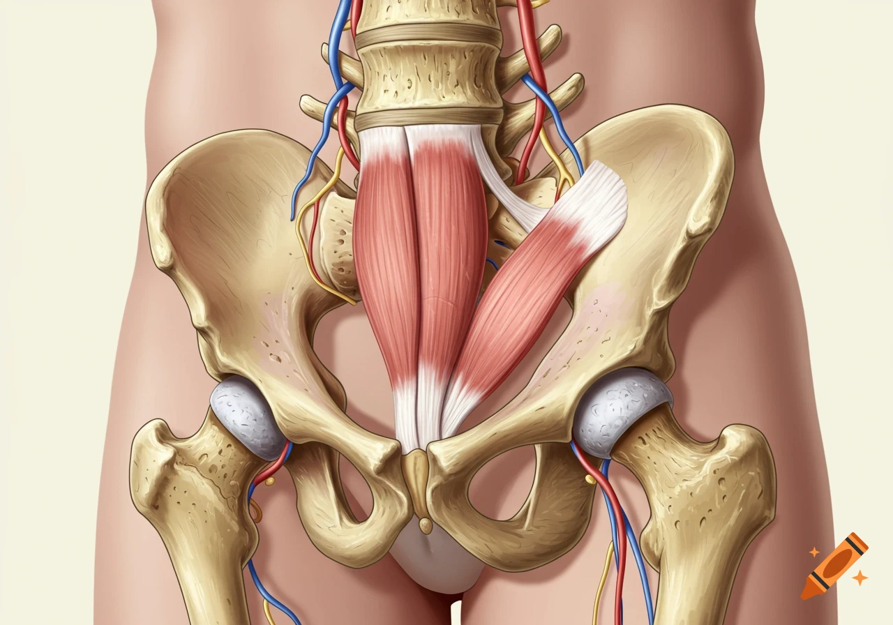
Detailed anatomical illustration of the human pelvis and lower back muscles, with the sacrum and a semi-transparent intergluteal cleft.

“Draw a detailed anatomical illustration from the posterior view of the human pelvis and lower back. Focus only on the tissues within and immediately around the intergluteal cleft (natal cleft), from the sacrum and coccyx (tailbone) down through the anal triangle of the perineum. Show the gluteus maximus muscles forming the outer buttocks, but highlight only the deep structures along the groove of the buttocks: the deep gluteal muscles (piriformis, gemelli, obturator internus, quadratus femoris), the sacrotuberous ligament, and the surrounding fascial layers. Render the intergluteal cleft and perineal midline as a clearly outlined, semi‑transparent strip, with these structures color‑coded and semi‑transparent, while the rest of the body is kept in a light gray or neutral medical‑style background to show only this region. Style: medical‑anatomy diagram, semi‑realistic, high‑detail, labeled layers optional, isometric posterior‑pelvis view, no genitals shown, no legs or abdomen beyond the hip bone and perineum.” Mehr sehen
More images like this