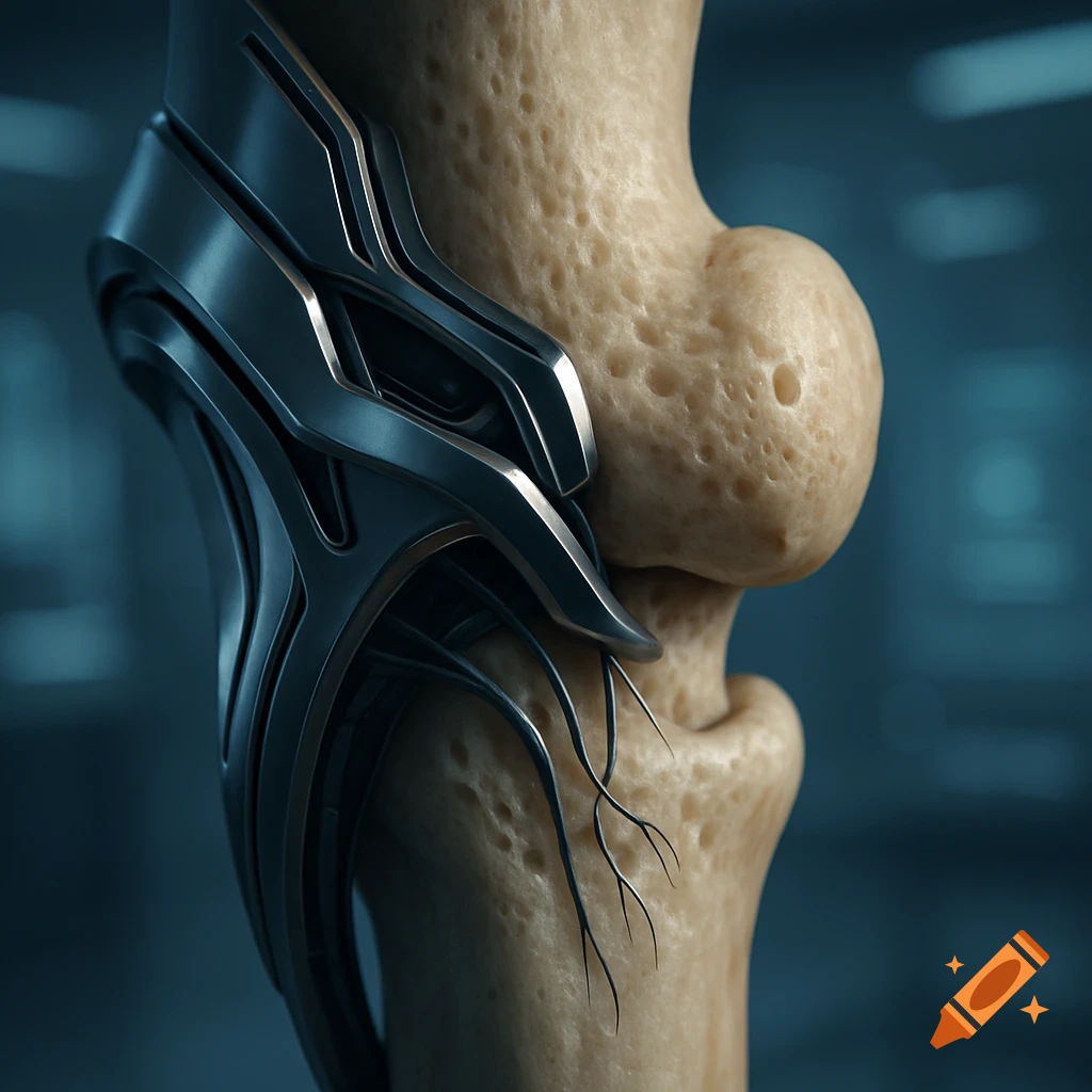
A detailed 3D scientific illustration shows a spherical bone being cut into a cube by a saw blade, revealing porous internal bone structure. Labeled diagram.

Scientific medical illustration of a porcine femoral head (pig femur head) in high detail. The image shows the spherical femoral head with a clearly visualized cubic sample being cut out from the center. Illustrate the cutting process step-by-step with subtle cut lines, semi-transparent slicing planes, and a removed cube section to show the internal bone structure. Emphasize the cutting geometry and the required saw cuts to extract a perfect cube from the bone. Clean textbook style, orthopedic and anatomical accuracy, neutral background, high clarity, labeled cutting planes, 3D-rendered scientific visualization, realistic bone texture, surgical precision aesthetic. Mehr sehen
More images like this